
 EN
EN
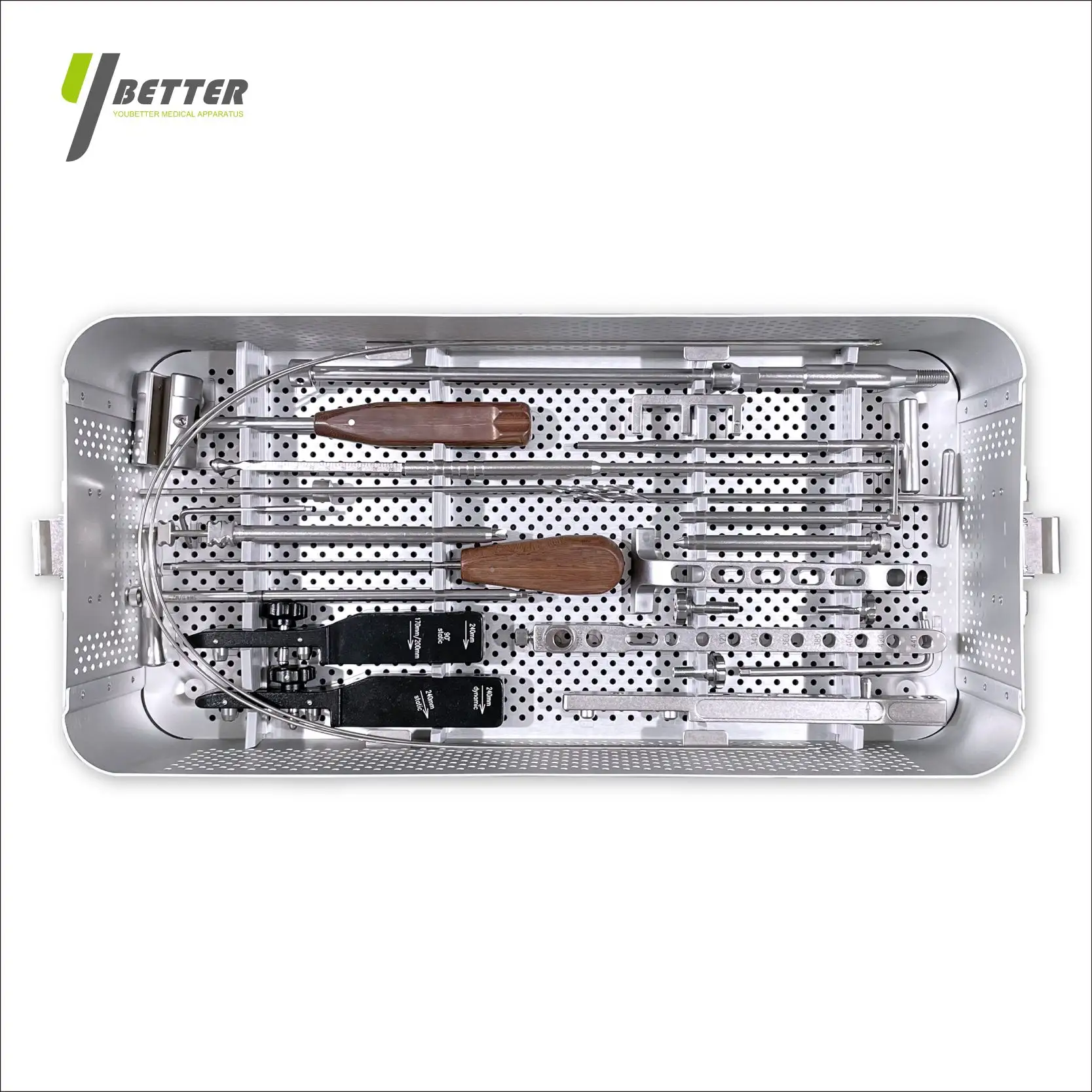
Atvērtie lūzumi ir viena no visgrūtākajām situācijām ortopēdiskās traumas pārvaldībā, kas prasa ātru lēmumu pieņemšanu un precīzu tehnisko izpildi. Iekšējās fiksācijas un ārējās stabilizācijas izvēle pamatā ietekmē pacienta iznākumus, inficēšanās risku un ilgtermiņa funkcionālo atveseļošanos. Ārējais fiksators... ārējais fiksators ir kļuvis par zelta standartu konkrētām pacientu populācijām un traumu veidiem, piedāvājot skaidras priekšrocības piesārņojuma kontroles, mīksto audu aizsardzības un posmu veidā veicamo ārstēšanas protokolu jomā. Ir būtiski saprast, kad izmantot ārējo fiksatoru salīdzinājumā ar citām fiksācijas metodēm traumas ķirurgiem un ortopēdiem, kuri pārvalda sarežģītas skeleta traumas.
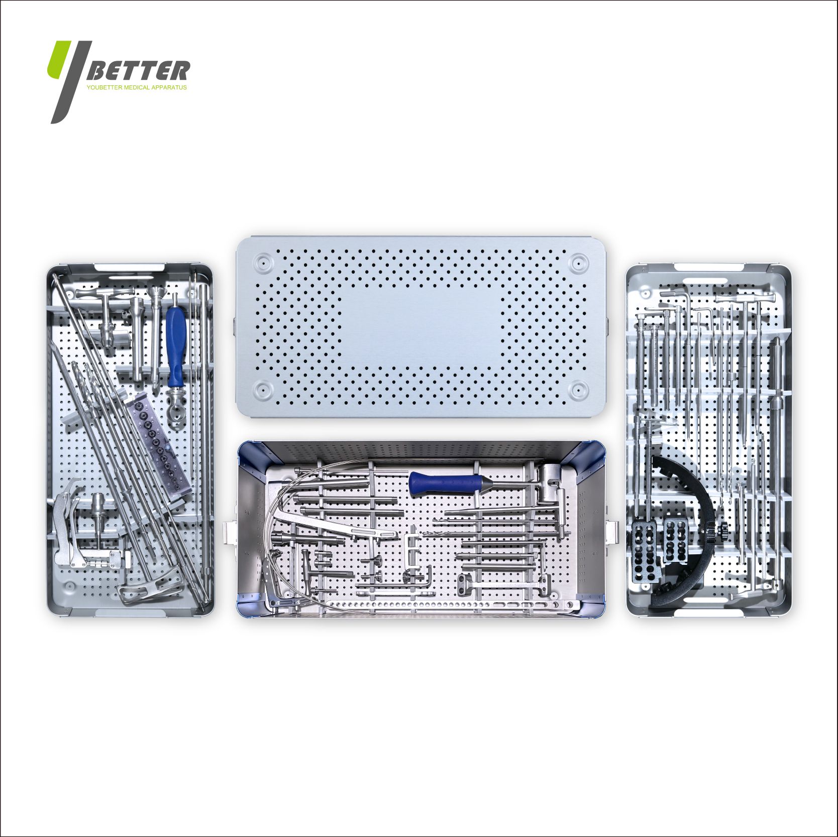
Ārējais fiksators ir būtisks instruments mūsdienu ortopēdiskās traumas ķirurģijā, nodrošinot nekavējoties skeleta stabilitāti, vienlaikus saglabājot traumas zonu, lai veiktu detalizētu mīksto audu novērtējumu un iespējamu galīgo rekonstrukciju. Šis pieejas veids pamatīgi atšķiras no iekšējās fiksācijas stratēģijām, kurām pirms metāla implantiem var droši novietot, nepieciešama sākotnēja brūces kontrole un asinsvadu integritāte. Ārējais fiksators ļauj ķirurgiem prioritāri veikt dzīvību glābjošas intervencijas, pārvaldīt sarežģītus piesārņojuma raksturus un atlikt galīgo fiksāciju līdz pacients sasniedz hemodinamisku stabilitāti un infekcijas risks ir pietiekami samazināts.
Klīniskās situācijas, kas veicina ārējā fiksatora izmantošanu
Gustilo-Anderson III pakāpes lūzumi un piesārņojuma raksturi
Gustilo–Anderson klasifikācija joprojām ir pamata rāmis, lai noteiktu, kad ārējais fiksators ir absolūti nepieciešams atvērtu lūzumu ārstēšanā. III pakāpes traumas, kas raksturīgas ar smagu mīksto audu zudumu, asinsritēs traucējumiem vai augsta ātruma mehānismiem, vienmēr prasa ārējā fiksatora izmantošanu, nevis nekavējoties iekšējo fiksāciju. Ārējais fiksators nodrošina neierobežotu piekļuvi secīgai brūces dezinficēšanai, ļaujot traumatoloģijas komandām noņemt dzīvību zaudējušos audus, izskalot piesārņotās zonas un novērtēt mikroasinsriti, neatraujot skeleta izvietojumu. III B un III C pakāpes lūzumos, kas saistīti ar spiediena traumām, šaušanas ievainojumiem vai sprādziena traumām, ārējais fiksators kļūst obligāts, jo mīksto audu bojājumi aptver daudz lielāku teritoriju nekā redzamā brūce, radot nezināmības zonas, kurām nepieciešama posmu veidā veikta izpēte un pakāpeniska mīksto audu rekonstrukcija pirms pastāvīgās metāla fiksācijas drošas piemērošanas.
Politraumas pacienti un bojājumu kontroles protokoli
Politraumas pacienti ar vairāku sistēmu traumām iegūst lielu labumu no ārējā fiksatora izmantošanas, jo šī fiksācijas metode prioritāri nodrošina ātru skeleta stabilizāciju, neprasa ilgu operācijas laiku. Kad traumas pacients parādās ar vienlaicīgām krūtiskās, vēdera vai galvas traumām, kas prasa steidzamu iejaukšanos, ārējais fiksators ļauj ortopēdiem sasniegt lūzuma redukciju un pagaidu stabilitāti minūtēs, nevis stundās. Šis traumas kontroles ortopēdiskais pieejas veids, izmantojot ārējo fiksatoru, samazina iekšoperācijas asins zudumu, minimizē šķidruma atjaunošanas nepieciešamību un paātrina pāreju uz galīgās aprūpes protokoliem. Īpaši lielu labumu no ārējā fiksatora sistēmām gūst pacienti ar nestabilām iegurņa lūzumiem kombinācijā ar garo kaulu traumām, jo šīs sistēmas nodrošina divzonu stabilizāciju, saglabājot hemodinamisko piekļuvi un ļaujot nepārtraukti monitorēt traumas stāvokli, neatkārtoti nemainot pacienta pozīciju operāciju galda virsmā.
Ārējo fiksācijas sistēmu tehniskās priekšrocības
Mīksto audu saglabāšana un infekciju profilakse
Ārējais fiksators mehāniski atdala traumas zonu no iekšējiem metāla implantiem, būtiski samazinot baktēriju kolonizāciju un biofilmu veidošanos gar lūzuma virsmām. Atšķirībā no iekšējās fiksācijas, kur metāla plāksnes un skrūves rada svešķermeņa robežvirknes, kas veicina infekcijas izveidošanos, ārējais fiksators ļauj nepārtraukti noplūdināt brūci un tieši uz lūzuma zonas lietot vietējos antiseptiskos līdzekļus. Infekciju biežums III pakāpes atvērtajos lūzumos, kurus ārstē ar ārējo fiksatoru sistēmām, paliek ievērojami zemāks nekā tiem, kurus ārstē ar nekavējoties veiktu iekšējo fiksāciju, jo īpaši tad, ja ārējā fiksatora stratēģiju kombinē ar piemērotu antibiotiku terapiju un secīgu dezinfekciju protokoliem. Ārējais fiksators arī ļauj vienkārši noņemt implanti, nebojājot sadzīvošo lūzuma vietu, tādējādi nodrošinot nevainojamu pāreju uz galīgo iekšējo fiksāciju, kad ir sasniegta mīksto audu pārklāšana un infekcijas risks ir novērsts posmu veidā veiktā rekonstrukcijā.
Regulējamība un progresīvās slodzes protokoli
Ārējās fiksācijas sistēmas nodrošina dinamisku pielāgojamību, ko stiprais iekšējais implantāts nevar atkārtot agrā ārstēšanas posmā. Kirurgi var mainīt samazināšanas leņķi, koriģēt rotācijas izkārtojumu un pielāgot saspiestās spējas visā ārstēšanas laikā, neiet operācijas telpā, lai veiktu oficiālas pārskatīšanas procedūras. Šī pielāgojamība ir ļoti vērtīga sarežģītajos lūzumu modeļos, kad sākotnējie samazināšanas lēmumi ir neoptimali vai ja nozīmīga mīkstās auduma pietūkums prasa pagaidu samazināšanas pielāgojumus. Ārējais fiksators ļauj arī progresīvu svaru nesēju protokolu, kas atbilst ārstēšanas bioloģijai, ļaujot kontrolēt mikromotīvu lūzumu vietās, kas paātrina kallu veidošanos, vienlaikus novēršot pārmērīgas sašķidrināšanas spējas, kas apdraud integrāciju. Pacienti ar ārējo fiksatoru lietošanu ievērojami ātrāk sasniedz funkcionālo atveseļošanās laiku, salīdzinot ar ilgstošu imobilizāciju kastēs vai ierobežojošiem iekšējiem fiksācijas ierīcēm, kas prasa ilgstošas bezsvaru fases.
Pacientu izvēles kritēriji un laikapstākļu apsvērumi
Sasites raksturlielumi, kas prasa ārējā fiksatora izmantošanu prioritāri
Konkrēti lūzumu raksturi un anatomiškās atrašanās vietas ļoti veicina ārējā fiksatora izmantošanu kā pirmo fiksācijas stratēģiju, nevis kā vēlāku pāreju uz citu metodi. Garo kaulu diaphīzes lūzumi ar smagu fragmentāciju, kaulu zudumu vai asinsvadu traumu parasti prasa ārējā fiksatora stabilizāciju, lai aizsargātu rekonstruētos asinsvadus un ļautu piekļuvi asinsvadu ķirurģijai posmu rekonstrukcijas laikā. Iegurņa lūzumiem ar hemodinamisku nestabilitāti absolūti nepieciešama ārējā fiksatora uzlikšana, jo šī metode nodrošina ātru stabilizāciju un iegurņa tilpuma samazināšanu, neveicot plašu operatīvu disekciju, kas pastiprina asiņošanu. Aizmugurējo kaulu lūzumi, kas saistīti ar gurna izvirzījuma traumām, bieži prasa ārējā fiksatora papildinājumu, lai saglabātu redukciju tajā laika posmā, kad mīksto audu pietūkums un asinsvadu kompromitācija neļauj izmantot tradicionālas ķirurģiskās fiksācijas metodes. Otrādi, vienkāršiem lūzumu raksturiem pacientiem ar minimālu mīksto audu traumu un lielisku asinsvadu stāvokli var tikt tieši pāriet uz iekšējo fiksāciju bez ārējā fiksatora starpposma, akcentējot, ka fiksācijas metodes izvēle ir atkarīga no integrētas novērtēšanas — ievainojuma smaguma, pacienta fizioloģijas un mīksto audu stāvokļa — nevis tikai no lūzuma atrašanās vietas.
Fiksācijas laiks un galīgās rekonstrukcijas logi
Ārējais fiksators parasti paliek uzstādīts septiņas līdz četrpadsmit dienas III pakāpes traumās, nodrošinot kritisku laika periodu, kurā mīkstās audu stāvoklis stabilizējas, infekcijas risks samazinās un var droši plānot galīgo rekonstrukciju. Šis posmu veidošanas pieeja ir diametrāli pretēja nekavējoties veiktajai iekšējai fiksācijai, kas prasa nevainojamu sākotnējo brūces apstrādi un ierobežo infekcijas risku līdz pieņemami zemām robežām. Ārējais fiksators ļauj traumatoloģijas komandām veikt secīgu brūces debridementu ik pēc divdesmit četrām līdz četrdesmit astoņām stundām, līdz brūce ir pilnībā attīrīta no kontaminācijas un visā lūzuma zonā ir apstiprināta mīksto audu dzīvotspēja. Kad šī iekaisuma fāze ir beigusies un mīksto audu segums kļūst iespējams, izmantojot vietējos vākus, ādas transplantāciju vai novēlotās rekonstrukcijas procedūras, ārējo fiksatoru var nomainīt pret galīgo iekšējo fiksāciju, kas optimizē ilgtermiņa funkcionālos rezultātus un ļauj īstenot paātrinātas reabilitācijas protokolus. Šī secīgā pieeja būtiski uzlabo infekciju brīvo sadzīšanas rādītājus un samazina amputācijas risku salīdzinājumā ar agresīvu primāro iekšējo fiksāciju piesārņotu traumu gadījumos.
Bieži uzdotie jautājumi
Kad ārējo fiksatoru vajadzētu pārvērst par iekšējo fiksāciju?
Pāreja no ārējā fiksatora uz iekšējo fiksāciju parasti notiek septītajā līdz divdesmit pirmajai dienai pēc traumas, kad ir samazinājusies mīksto audu pietūsne, infekcijas risks ir pietiekami samazināts ar atkārtotām debridement procedūrām un ir apstiprināti galīgie plāni mīksto audu rekonstrukcijai. Ārējais fiksators jāaizvieto ar iekšējo fiksāciju tad, kad lūzuma zonā ir skaidri redzama robeža starp dzīvo un nevieglo audu, bakterioloģisko kultūru rezultāti apstiprina kontaminācijas kontroli un pacients sasniedz hemodinamisku stabilitāti, kas ļauj veikt ilgākas operācijas. Vēlāka pāreja pēc trīs nedēļām var pasliktināt sadzīšanas rezultātus, ļaujot sekundārai infekcijai izveidoties vai pārmērīgiem kaula audu veidošanās procesiem, kas sarežģī turpmākās iekšējās fiksācijas metodes. Agrīna pāreja pirms pietiekamas mīksto audu stabilizācijas rada risku atkārtotai kontaminācijai un infekcijas pasliktināšanai, tādēļ fiksācijas metodes maiņai jānotiek precīzi saskaņā ar bioloģiskajiem sadzīšanas principiem, nevis patvaļīgi balstoties uz kalendāra termiņiem.
Vai ārējie fiksatori ir piemēroti visiem atvērtajiem kaulu lūzumiem?
Izplešu I. un II. pakāpes atvērtas lūzumi ar minimālu mīksto audu zudumu un zemu inficēšanās risku var tikt tieši stabilizēti ar iekšējo fiksāciju, neizmantojot ārējo fiksatoru kā starpposmu, ja pacienta fizioloģiskais stāvoklis ļauj ilgstošām operācijām un inficēšanu iespējams pilnībā novērst pirmajā operācijā. Tomēr daudzi traumas ķirurgi ieteic ārēja fiksatora izmantošanu II. pakāpes traumās ar būtisku mīksto audu bojājumu vai asinsritēs traucējumiem, atzīstot, ka ārēja fiksatora uzlikšanai nepieciešamais nelielais papildu operācijas laiks ir ievērojami pārsvars salīdzinājumā ar labāku infekciju kontroli un uzlabotu stadiju sadalījuma elastību. III. pakāpes traumas vienmēr prasa ārēja fiksatora izmantošanu, jo pastāv smags mīksto audu zudums, asinsritēs traucējumi vai inficēšanas raksturs, kas neļauj droši veikt iekšējo fiksāciju akūtā traumas stadijā. Ārējais fiksators kalpo kā pamata stabilizācijas stratēģija visos inficētos traumu gadījumos, ļaujot veikt turpmākus rekonstrukcijas lēmumus, kas būtu neiespējami, ja iekšējā fiksācija būtu veikta pāragri.
Kā ārējā fiksatora lietošana ietekmē ilgtermiņa funkcionālos rezultātus?
Ārējā fiksatora lietošana pienācīgi atlasītajiem atklāta lūzuma pacientiem nodrošina augstāku ilgtermiņa funkcionālo rezultātu salīdzinājumā ar agresīvām primārajām iekšējās fiksācijas stratēģijām, galvenokārt, samazinot infekciju skaitu, samazinot amputācijas risku un paātrinot rehabilitācijas termiņus. Pacientiem, kuriem tika veikta pakāpeniska ārējā fiksatora fiksācija, kam sekoja aizkavēta iekšējā fiksācija, novērota uzlabojusies kustības jomas, uzlabojusies spēka atjaunošanās un uzlabojās darba devēju atgriešanās termiņi salīdzinājumā ar pacientiem, kuriem pēc neveiksmīgas primārās iekšējās fiksācijas mēģinājumu Ārējā fiksatora regulējamais stāvoklis ļauj optimāli izvietot lūzumu, neskarot mīkstās audus, samazinot komplikācijas un mehanisku disfunkciju, kas rodas pēc gadiem pēc sākotnējās lūzuma ārstēšanas. Tomēr ārējais fiksators pagarina ārstēšanas ilgumu un palielina pacientu nepatīkamas sajūtas stabilizācijas posmā, kas prasa rūpīgu konsultāciju attiecībā uz gaidāmo laiku un reabilitācijas prasībām, bet uzsver, ka šī pakāpeniska pieeja prioritē infekciju profilaksi un ekstremitātēm saglabāšanu, nevis īstermiņa ērta rīcība.