
The evolution of surgical medicine has brought forward one of the most significant shifts in patient care: the widespread adoption of minimally invasive surgery. At the heart of this transformation lies the role of orthopedic implants, which have been engineered not only to restore function and structural integrity to the musculoskeletal system, but also to do so through the smallest possible disruption to surrounding tissue. Understanding how orthopedic implants contribute to minimally invasive surgery requires a closer look at their design principles, material innovations, and the procedural workflows they enable.
For surgical teams and hospital procurement specialists alike, the relationship between orthopedic implants and minimally invasive techniques is not merely academic. It directly impacts patient recovery times, complication rates, hospital stays, and overall clinical outcomes. As demand for less invasive procedures grows across spine, joint, and trauma surgery, the design and selection of orthopedic implants have become critical decisions that shape every stage of the surgical process — from planning through rehabilitation.
The Design Philosophy Behind MIS-Compatible Orthopedic Implants
Profile Reduction and Low-Profile Architecture
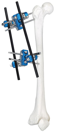
One of the foundational ways orthopedic implants contribute to minimally invasive surgery is through their physical profile. Traditional implants were designed for open surgery, where large incisions provided ample exposure. In contrast, modern orthopedic implants intended for MIS procedures are engineered with low-profile geometries that can be introduced through narrow portals, cannulas, or tubes without requiring extensive tissue retraction.
Low-profile design means that screws, rods, plates, and cages can be delivered and seated without displacing large volumes of soft tissue. This is especially critical in spinal surgery, where the paraspinal musculature must be preserved to ensure post-operative strength and stability. The dimensional precision required of these orthopedic implants demands advanced machining tolerances and material choices that support both miniaturization and load-bearing capacity simultaneously.
Engineers designing orthopedic implants for MIS applications must balance competing demands: the implant must be small enough to pass through a limited access corridor, yet robust enough to perform its biomechanical function under physiological loading conditions. This challenge has driven significant innovation in implant geometry, surface finish, and fixation mechanism design.
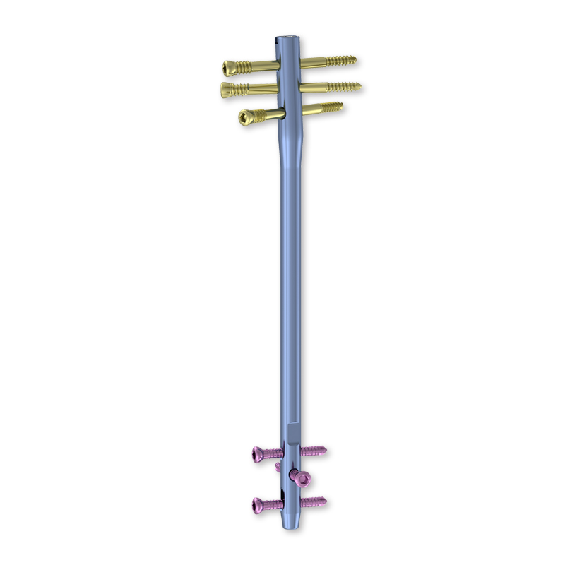
Modular and Expandable Implant Systems
Another key contribution of orthopedic implants to minimally invasive surgery is the rise of modular and expandable systems. Rather than inserting a fully assembled, rigid structure through a small incision — which would require that incision to be as large as the implant — surgeons can now insert components in a collapsed or disassembled state and expand or lock them in place once properly positioned.
Expandable interbody cages used in spinal fusion procedures are a prime example. These orthopedic implants are introduced at a reduced height and then expanded within the disc space to restore proper segmental height and lordosis. This approach allows the surgeon to work through a minimally invasive corridor while still achieving the biomechanical result previously only possible through open surgery.
Modular systems also reduce the number of components that must be individually introduced, decreasing operative time and the mechanical complexity of the MIS procedure. For procurement teams, this modularity translates into streamlined instrument and implant sets that are easier to sterilize, manage, and track across procedures.
Material Science and Its Role in MIS Implant Performance
Titanium Alloys and Their MIS Advantages
The materials used in orthopedic implants have a direct bearing on how well they perform in minimally invasive surgical contexts. Titanium alloys remain among the most widely used materials for orthopedic implants due to their excellent strength-to-weight ratio, biocompatibility, and radiolucency under fluoroscopic and CT imaging — all properties that are particularly valuable in MIS settings.
In minimally invasive procedures, surgeons rely heavily on intraoperative imaging to confirm implant positioning without direct visual access to the surgical site. Orthopedic implants made from titanium alloys produce minimal imaging artifact, allowing surgeons to verify placement accurately through fluoroscopy or navigation systems. This imaging compatibility is not incidental — it is a fundamental design requirement for orthopedic implants used in MIS.
The osseointegration properties of titanium also support long-term fixation without requiring the extended healing periods associated with less biocompatible materials. In MIS procedures where the healing environment is already optimized by reduced soft tissue disruption, titanium orthopedic implants accelerate the overall biological recovery process.
PEEK and Advanced Polymer Composites
Polyetheretherketone, commonly known as PEEK, has emerged as another material of significant importance for orthopedic implants in minimally invasive surgery. PEEK offers an elastic modulus closer to cortical bone than metal, which reduces the risk of stress shielding — a condition where the implant bears too much load and the adjacent bone weakens due to insufficient mechanical stimulation.
For spinal orthopedic implants in particular, PEEK interbody devices allow for clear visualization of fusion progress on postoperative imaging because they do not produce the metal artifact that can obscure assessment. This is clinically valuable when evaluating outcomes through MRI or CT following minimally invasive spinal fusion procedures.
Advanced composites that combine PEEK with carbon fiber or hydroxyapatite surface treatments are pushing the boundaries further. These hybrid orthopedic implants retain the imaging benefits and biomechanical properties of PEEK while improving biological integration. For hospitals investing in MIS programs, understanding these material distinctions is essential to selecting orthopedic implants that align with both procedural requirements and patient outcome goals.
Instrumentation Systems That Enable Implant Delivery in MIS
Purpose-Built MIS Instrument Sets
Orthopedic implants cannot be evaluated in isolation from the instruments required to deliver them. In minimally invasive surgery, the instrumentation system is just as critical as the implant itself. Dedicated orthopedic implants delivery systems have been developed to allow percutaneous or tubular access, precise trajectory control, and secure fixation — all while operating within the spatial constraints of a minimally invasive corridor.
MIS instrument sets for spinal procedures, for example, typically include cannulated screwdrivers, extended-handle reducers, and rod delivery systems that allow the surgeon to manipulate implant components from outside the patient's body while targeting deep spinal anatomy through small skin incisions. The design of these instruments must be ergonomically aligned with the implants they serve, ensuring reliable engagement without slippage or misalignment.
For procurement and supply chain teams, sourcing orthopedic implants alongside their corresponding MIS instrument sets as integrated systems reduces compatibility risks and ensures that the surgical team has everything needed for efficient, safe implant delivery. The instrument set is not an accessory — it is a co-dependent component of the MIS implant system.
Navigation and Robotic Assistance in Implant Placement
Surgical navigation and robotics have become increasingly intertwined with the use of orthopedic implants in minimally invasive procedures. These technologies compensate for the reduced direct visualization inherent in MIS by providing real-time guidance that helps surgeons place orthopedic implants with high accuracy despite the limited operative field.
Navigation systems use preoperative imaging data — typically CT scans — to create a virtual surgical map, enabling the placement of pedicle screws, acetabular cups, or femoral stems with millimeter-level precision. Orthopedic implants designed for navigation-assisted placement often incorporate reference features or registration markers that integrate with tracking systems used intraoperatively.
Robotic arms take this a step further by physically constraining the instrument trajectory within a predefined safe zone. This is particularly important when placing orthopedic implants near critical neurovascular structures, where even minor deviations in a minimally invasive approach could have serious consequences. The convergence of advanced orthopedic implants with navigation and robotics is one of the most powerful accelerators of MIS adoption in orthopedic surgery today.
Clinical Outcomes and Patient Benefits Driven by MIS Implant Integration
Reduced Tissue Trauma and Faster Recovery
The most direct patient benefit tied to the integration of orthopedic implants with minimally invasive surgical techniques is the dramatic reduction in tissue trauma. When orthopedic implants are designed specifically for MIS delivery, surgeons can achieve the same stabilization or reconstruction objectives as open surgery while preserving the muscles, ligaments, and soft tissue structures that surround the operative site.
This tissue preservation translates clinically into reduced postoperative pain, lower blood loss, decreased need for transfusion, and significantly shorter hospital stays. Patients who receive orthopedic implants through minimally invasive approaches consistently report faster returns to daily activities and improved satisfaction scores compared to those who undergo traditional open procedures with equivalent implant goals.
For healthcare systems operating under value-based care models, these outcomes represent both clinical and economic advantages. Reduced complications and shorter admissions lower the cost per episode of care, making the case for investing in the right orthopedic implants and supporting MIS infrastructure.
Long-Term Fixation Integrity and Bone Preservation
Beyond the immediate perioperative benefits, orthopedic implants contribute to minimally invasive surgery by supporting better long-term outcomes through enhanced bone preservation. MIS approaches inherently disturb less periosteum and vascularity around bone, which improves the local biological environment for implant integration and fusion.
When orthopedic implants are placed through minimally invasive corridors, the surrounding bone retains more of its native blood supply, accelerating healing and reducing the risk of implant loosening or non-union. This is particularly important in spinal fusion, where the long-term stability of the construct depends on successful osseointegration between the orthopedic implants and the adjacent vertebral endplates.
Orthopedic implants with textured or porous surface features further enhance this integration by encouraging bone ingrowth at the implant-bone interface. These surface engineering strategies are most effective precisely when the MIS approach has preserved the biological environment that supports such ingrowth — making the implant design and the surgical technique genuinely synergistic.
FAQ
What types of orthopedic implants are most commonly used in minimally invasive spine surgery?
The most commonly used orthopedic implants in minimally invasive spine surgery include percutaneous pedicle screw systems, expandable interbody cages, and lateral lumbar interbody fusion devices. These implants are specifically designed for introduction through small incisions or tubular retractors, and they are often paired with dedicated MIS instrument sets that allow the surgeon to achieve proper implant positioning without open exposure of the spine.
How do orthopedic implants support imaging guidance during minimally invasive procedures?
Orthopedic implants used in MIS procedures are typically manufactured from materials such as titanium or PEEK that produce minimal artifact on fluoroscopic and CT imaging. This radiolucent or artifact-reducing property is essential because surgeons in minimally invasive surgery rely on real-time imaging rather than direct vision to confirm implant placement. Some orthopedic implants also incorporate registration features that interface with surgical navigation systems for enhanced accuracy.
Are orthopedic implants designed for MIS procedures as durable as those used in open surgery?
Yes. Orthopedic implants designed for minimally invasive surgery undergo the same rigorous biomechanical testing and regulatory review as those used in open procedures. Their reduced physical profile does not compromise structural integrity because engineers account for loading conditions when designing MIS-compatible orthopedic implants. In many cases, the preservation of surrounding musculature and vascularity achieved through MIS actually improves the long-term performance environment of the implant.
What should hospitals consider when procuring orthopedic implants for an MIS surgical program?
Hospitals building or expanding an MIS surgical program should consider orthopedic implants in the context of complete system compatibility — meaning the implants, instrumentation, and imaging or navigation support must be designed to work together. Procurement teams should evaluate the modularity of the implant system, the availability of dedicated MIS instrument sets, surgeon training support, and the implant manufacturer's clinical evidence base. Selecting orthopedic implants that are optimized for the specific MIS procedures being performed is essential to achieving consistent and reproducible clinical outcomes.
Table of Contents
- The Design Philosophy Behind MIS-Compatible Orthopedic Implants
- Material Science and Its Role in MIS Implant Performance
- Instrumentation Systems That Enable Implant Delivery in MIS
- Clinical Outcomes and Patient Benefits Driven by MIS Implant Integration
-
FAQ
- What types of orthopedic implants are most commonly used in minimally invasive spine surgery?
- How do orthopedic implants support imaging guidance during minimally invasive procedures?
- Are orthopedic implants designed for MIS procedures as durable as those used in open surgery?
- What should hospitals consider when procuring orthopedic implants for an MIS surgical program?