
 EN
Evaluating a cannulated screw in osteoporotic bone means assessing more than whether the implant appears seated. Lower bone density can reduce the purchase available to the threads, so stability depends on the fracture pattern, placement, construct, and patient factors together. A cannulated screw should be judged against the intended fixation goals, not by a single image or insertion sensation.
A useful assessment combines preoperative planning, intraoperative checks, and follow-up review. For a cannulated screw, clinicians consider whether the trajectory matches the anatomy, whether the threads engage the intended bone, and whether the construct controls expected forces. These observations help identify concerns early, while recognizing that imaging and clinical judgment cannot guarantee a particular healing outcome.
Assessing fixation with imaging and examination
Reviewing position and bone engagement
Start by confirming the fracture site, reduction, and planned load-sharing role of the implant. On appropriate views, a cannulated screw should follow the intended path without entering a joint or compromising nearby structures. Assess whether its threaded portion engages the planned region of bone; a cannulated screw placed accurately may still have limited purchase where bone quality is poor.
Review more than one imaging view because a single projection can conceal malposition, cortical breach, or an unintended gap. Compare the screw trajectory with the fracture line and surrounding anatomy, and look for loss of reduction that may suggest inadequate control. A cannulated screw assessment should document both satisfactory features and uncertainties, rather than treating apparent alignment as proof of secure fixation.
Interpreting intraoperative and follow-up findings
During insertion, the operator may note resistance, depth, and the relationship between the implant and prepared path, but tactile feedback is only one part of evaluation. Osteoporotic bone can offer less predictable resistance, so a cannulated screw should not be considered stable solely because insertion felt firm. Use the planned imaging checks and operative assessment to judge the construct as a whole.
After fixation, follow-up images can be compared with the immediate postoperative baseline for changes in alignment, implant position, or fracture gap. The meaning of a finding depends on anatomy, healing stage, symptoms, and the intended loading plan. If a cannulated screw appears to migrate or reduction changes, the treating team can assess whether this reflects a stability concern and adjust monitoring or management.
Factors that influence stability in low-density bone
Fracture geometry and screw trajectory
Fracture configuration affects the forces the fixation must resist. Fragment size, fracture orientation, comminution, and the quality of reduction all shape how a cannulated screw can contribute to stability. A trajectory should be planned around the target anatomy and fracture mechanics; simply increasing screw length or changing direction does not automatically improve purchase in osteoporotic bone.
Consider whether the proposed cannulated screw can control rotation, shear, and displacement for the specific fracture. The number and arrangement of fixation elements, where relevant, depend on the clinical plan and anatomical constraints. Poor reduction can concentrate stress at the implant-bone interface, so a cannulated screw cannot compensate for an unaddressed fracture gap or unsuitable construct design.
Bone quality, implant fit, and patient demands
Bone density is important, but local bone quality may vary within the same region. Prior imaging, the patient’s history, and operative observations can inform expectations about a cannulated screw’s purchase. Thread engagement, fit, and the intended endpoint should be considered together, with care not to infer dependable holding strength from a general diagnosis of osteoporosis alone.
Stability is also affected by forces placed on the fixation during daily activity and rehabilitation. Body mechanics, mobility, fall risk, and the planned weight-bearing progression can influence construct demands. A cannulated screw evaluation therefore connects implant placement to a realistic loading plan, rather than assuming the same fixation will behave identically in every patient or clinical setting.
Applying a structured evaluation to clinical decisions
Linking the assessment to the fixation goal
Before judging stability, clarify the goal: maintaining reduction, limiting rotation, supporting a fragment, or enabling a defined rehabilitation pathway. Then assess whether the cannulated screw position and construct address that goal without creating avoidable anatomical risk. This framing makes the review specific and helps the team distinguish a technical concern from a feature that is acceptable for the treatment plan.
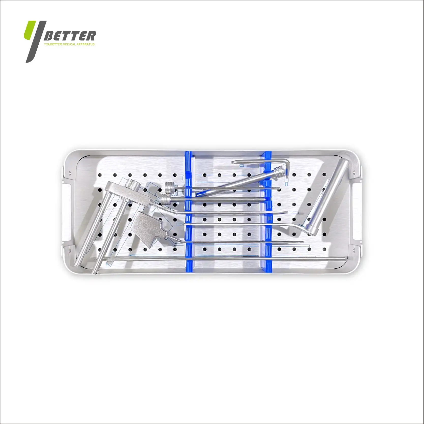
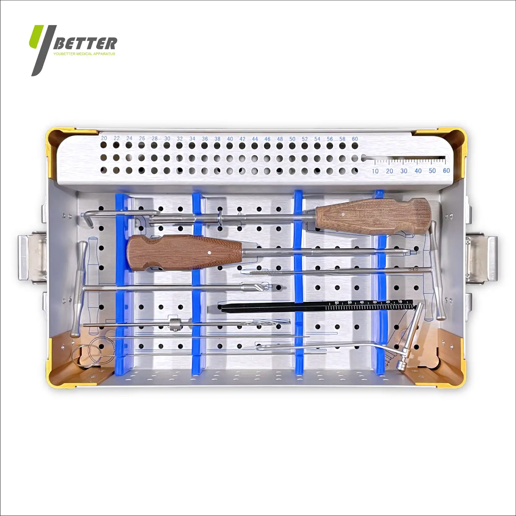
A practical review brings together reduction quality, imaging views, bone engagement, fracture pattern, and expected loading. Record what is known, what remains uncertain, and which findings would prompt reassessment. When relevant, the cannulated screw instrumentation and preparation workflow should also be considered in relation to the planned technique, without treating equipment alone as evidence of stability.
Monitoring concerns and adjusting the plan
If assessment raises concern, first identify the specific issue: reduction, trajectory, thread purchase, construct mechanics, or patient loading. The response depends on the clinical context and may involve additional review, closer follow-up, or a change in management by the treating team. A cannulated screw finding should be interpreted alongside symptoms and the full imaging record, not in isolation.
Consistent documentation makes serial evaluation more useful. Note the views obtained, implant position, fracture alignment, and any interval changes, then relate them to symptoms and rehabilitation status. This process does not remove uncertainty in osteoporotic bone, but it supports reasoned decisions about a cannulated screw and makes the basis for continued observation or further action clear.
FAQ
Can imaging alone confirm cannulated screw stability?
No single image proves stability. Imaging shows position and alignment, while interpretation also considers fracture mechanics, bone quality, symptoms, and loading. A cannulated screw should be evaluated using suitable views and, when available, comparison over time. The treating team integrates those findings with operative details and clinical examination.
Which findings may raise concern after fixation?
Potential concerns include interval loss of reduction, a change in implant position, an unexpected fracture gap, or symptoms that warrant reassessment. These findings do not establish a cause by themselves. Review a cannulated screw in context, comparing current images with baseline and considering healing stage, activity, and the original fixation goal.
Does osteoporosis always mean a cannulated screw will be unstable?
No. Osteoporosis can make fixation assessment more demanding, but stability depends on several connected factors, including fracture pattern, reduction, placement, and loading. A cannulated screw should be assessed individually rather than judged by diagnosis alone. The clinical team can explain how these factors inform the patient’s follow-up and activity plan.