
 EN
EN
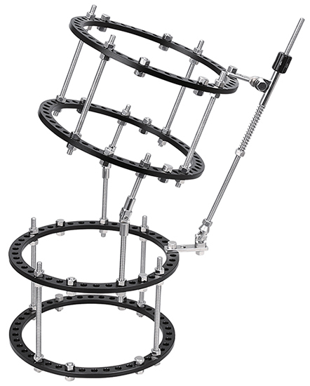
Combining an external fixator with internal fixation represents one of the most sophisticated approaches in modern orthopedic trauma surgery. This hybrid technique leverages the strengths of both fixation methods to address complex bone fractures that would be difficult or impossible to manage using either approach alone. Surgeons increasingly turn to this combined strategy when treating severe fractures, particularly those involving multiple fragments, significant soft tissue damage, or unstable bone patterns that demand both stability and flexibility during healing.
Yes, an external fixator can absolutely be combined with internal fixation devices to create a hybrid fixation construct that maximizes biomechanical support and clinical outcomes. The decision to use both methods together depends on fracture complexity, anatomical location, soft tissue condition, and the surgical timeline. Understanding when and how to integrate these techniques is essential for trauma surgeons seeking optimal patient results.
Understanding Hybrid Fixation with External Fixator and Internal Methods
The Fundamentals of Combined Fixation Approaches
An external fixator works by stabilizing bone fragments through percutaneous pins or half-pins that connect to an external frame, while internal fixation uses plates, screws, and intramedullary devices placed directly within or on the bone. When surgeons combine these approaches, the external fixator provides immediate provisional stability and load-sharing capability, while the internal fixation—often represented by plates or screws—offers anatomical reduction and long-term structural support. This hybrid strategy is particularly valuable in polytrauma situations where rapid stabilization is critical yet anatomical precision is equally important. The external fixator can be applied quickly in the emergency department or operating room, allowing surgeons to stabilize the limb and manage soft tissue swelling before undertaking more definitive internal fixation. Simultaneously, the internal fixation ensures that once the initial trauma phase subsides, the bone fragments remain precisely aligned for optimal healing and function.
Clinical Scenarios Where Combined External Fixator and Internal Fixation Proves Essential
The most compelling use cases for combining an external fixator with internal fixation involve complex periarticular fractures, such as pilon fractures of the distal tibia or proximal humerus fractures with multiple fragments. In pilon fractures, for instance, surgeons may apply an external fixator across the ankle joint as a temporary measure to restore limb length and alignment, then perform definitive open reduction and internal fixation of the tibial plafond once soft tissue swelling resolves. Another important scenario includes segmental fractures where the external fixator maintains overall limb length and axis while internal fixation devices address the specific fracture lines. High-energy pelvic fractures often benefit from temporary external fixator placement followed by plate and screw fixation, especially when the fracture pattern threatens pelvic stability or vascular structures. The external fixator acts as a 'holding device,' keeping the pelvis reduced while allowing safer surgical access for internal fixation placement.
Biomechanical and Clinical Benefits of Hybrid External Fixator and Internal Fixation Constructs
Load-Sharing and Stress Distribution in Hybrid External Fixator Systems
One of the primary advantages of combining an external fixator with internal fixation is enhanced load-sharing across the entire fracture site. The external fixator provides dynamic off-loading through its frame geometry, allowing micromotion that stimulates callus formation and promotes secondary bone healing. Simultaneously, the internal fixation—typically a locking plate or compression device—bears a portion of the load, creating a redundant support system that reduces the risk of fixation failure. This dual-load architecture is biomechanically superior to either method alone in many complex fracture patterns. When the external fixator is properly tensioned and configured, it can reduce the bending moment across the fracture site by up to 40 percent, thereby decreasing the stress borne by internal fixation devices. This is particularly beneficial in osteoporotic bone or in revision cases where the external fixator compensates for potentially compromised screw purchase in weak bone stock. The combination also permits earlier weight-bearing in some fracture patterns, as the redundancy allows surgeons to progress rehabilitation more aggressively without risking construct failure.
Soft Tissue Management and Infection Prevention Through External Fixator Integration
A critical clinical benefit of using an external fixator alongside internal fixation is superior soft tissue management during the acute post-trauma phase. Severe fractures are frequently accompanied by soft tissue injury, compartment syndrome risk, and vascular compromise. Applying an external fixator first allows immediate stabilization without the time commitment of internal fixation surgery, thereby reducing tissue damage from fracture fragment motion. This 'damage control' approach aligns perfectly with modern trauma protocols emphasizing early stabilization over early definitive fixation. Once swelling subsides—typically within three to seven days—surgeons can proceed with internal fixation using smaller incisions or minimally invasive techniques, as the external fixator maintains provisional alignment. This staged approach significantly reduces infection risk, particularly in open fractures, because the external fixator permits easier wound care, prevents bacterial biofilm formation on internal hardware during the contaminated phase, and allows surgeons to assess soft tissue viability before committing to extensive internal fixation. Studies demonstrate that hybrid external fixator and internal fixation constructs show lower infection rates than either method alone in high-energy pilon fractures and open femur fractures.
Surgical Technique, Timing, and Practical Implementation of External Fixator with Internal Fixation
Sequencing and Frame Configuration for External Fixator Placement
The surgical sequence when combining an external fixator with internal fixation requires careful planning and typically follows a 'provisional then definitive' model. The external fixator is usually applied first, often under general anesthesia or even in a resuscitation setting during massive trauma cases. The surgeon positions percutaneous half-pins or pins in safe zones—areas where pin placement avoids nerves, blood vessels, and zones where internal fixation will later be applied. For femur fractures, the external fixator frame typically bridges the fracture, with proximal pins above the hip and distal pins below the knee, maintaining anatomical length and alignment. For tibia fractures, a similar approach applies, with the frame positioned on the lateral aspect to avoid the anterior tibial compartment. Once the patient stabilizes and soft tissue swelling plateaus, the surgeon proceeds with internal fixation, often removing the external fixator frame after definitive hardware is secured. However, in some complex cases—particularly in elderly patients with compromised bone quality or revision scenarios—surgeons may leave the external fixator in place for four to six weeks, creating a truly hybrid construct that provides redundant fixation during the critical early healing phase.
Integration with Internal Fixation Devices and Hardware Considerations
When the external fixator remains in place during internal fixation, the specific choice of internal fixation hardware matters significantly. Locking compression plates are often preferred because they provide fixed-angle fixation that does not rely on friction between the plate and bone, making them ideal for use alongside an external fixator that partially load-shares. Intramedullary nailing is another excellent option when the external fixator is configured on the lateral cortex, allowing unobstructed nail insertion along the medullary canal. Surgeons must ensure that pin sites for the external fixator do not conflict with planned screw corridors for plate fixation. This typically means positioning pins in 'safe zones' such as the proximal femur lateral cortex or proximal tibia anterolateral cortex, zones well away from the primary plate fixation sites. The external fixator frame's geometry should also allow adequate working space for the surgical team, so the frame may be partially disassembled or repositioned during internal fixation implantation. Once internal fixation is complete and radiographs confirm anatomical alignment and hardware position, the external fixator can typically be removed if sufficient stability is achieved through internal fixation alone.
Common Challenges and Best Practices in External Fixator and Internal Fixation Management
Addressing Pin Tract Complications and Frame Stability
Even though the external fixator is often temporary when combined with internal fixation, pin tract infections remain a practical concern. Modern best practices emphasize pin care protocols from the moment the external fixator is applied, including regular cleaning with antiseptic solution and sterile technique during pin site management. Surgeons should assess pin loosening regularly; a loose pin may reduce the external fixator's contribution to overall construct stability. When removing the external fixator after internal fixation is secured, surgeons must do so systematically, typically removing one half-pin at a time and reassessing radiographic alignment after each removal to confirm that the internal fixation alone maintains adequate reduction. In some challenging cases, particularly in osteoporotic bone or revision surgeries, a staged approach may leave the external fixator in place for four to eight weeks even after internal fixation is complete, allowing the external fixator to support healing during the critical phase when callus formation is most active.
Balancing Early Mobilization with Construct Protection
A key challenge when using hybrid external fixator and internal fixation is determining the appropriate weight-bearing and mobilization timeline. The presence of an external fixator provides additional stability that may permit earlier weight-bearing than internal fixation alone, yet the fracture's inherent stability and the patient's bone quality remain the limiting factors. Surgeons must communicate clearly with the rehabilitation team about the biomechanical contribution of both devices. In many cases, partial weight-bearing may begin within days of external fixator application, with progression to full weight-bearing once internal fixation is secure and soft tissue healing is progressing well. The hybrid construct allows this flexibility because either the external fixator or the internal fixation can be adjusted or modified if early mobilization reveals inadequate stability. Regular clinical and radiographic follow-up is essential during the first four to six weeks to confirm that the construct is performing as intended and that bone healing is progressing normally.
FAQ
Is it always necessary to use both an external fixator and internal fixation together?
No, combining an external fixator with internal fixation is reserved for specific fracture patterns and clinical scenarios rather than routine cases. Simple fractures with minimal soft tissue injury typically respond well to internal fixation alone, while some highly comminuted fractures in good bone quality may be managed adequately with an external fixator alone. The decision hinges on fracture complexity, soft tissue condition, patient stability, and anatomical location. Surgeons use the hybrid approach when the clinical situation demands the unique benefits that each method provides—the external fixator's capacity for rapid stabilization and soft tissue management combined with the internal fixation's anatomical precision and long-term structural support.
How long does an external fixator typically remain in place when used with internal fixation?
The duration varies depending on the fracture pattern and clinical progression. In many trauma cases, the external fixator may be removed within one to two weeks after internal fixation is complete, once imaging confirms adequate reduction and stable hardware placement. In more complex cases or those involving poor bone quality, the external fixator may remain for four to eight weeks, providing hybrid support during the critical early healing phase when callus formation is most active. Pin sites are managed continuously during the external fixator's presence, and removal is performed systematically with radiographic confirmation that the internal fixation alone maintains alignment.
What are the main risks of combining an external fixator with internal fixation?
The primary risks include pin tract infections from the external fixator, potential hardware infection if soft tissue contamination occurs during the transition period, and increased surgical time and cost compared to single-method fixation. Additionally, the external fixator's pins must be carefully positioned to avoid conflicting with internal fixation corridors, requiring detailed preoperative planning. Patient compliance with pin care protocols is essential to minimize infection risk. However, when properly executed, the benefits of the hybrid approach typically outweigh these risks in complex fracture scenarios where either method alone would be insufficient.
Table of Contents
- Understanding Hybrid Fixation with External Fixator and Internal Methods
- Biomechanical and Clinical Benefits of Hybrid External Fixator and Internal Fixation Constructs
- Surgical Technique, Timing, and Practical Implementation of External Fixator with Internal Fixation
- Common Challenges and Best Practices in External Fixator and Internal Fixation Management
- FAQ