
 EN
EN
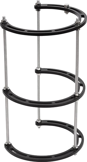
An external fixator is a critical orthopedic device used to stabilize fractured bones and promote proper alignment during the healing process. Determining how long an external fixator should remain in place depends on multiple factors including fracture type, location, severity, and the patient's individual healing response. Understanding the appropriate duration for external fixator retention is essential for optimizing patient outcomes and preventing complications such as infection, loss of alignment, or prolonged disability. Medical professionals must balance the need for adequate bone stability with the risks associated with prolonged external fixator use.
The duration an external fixator remains in place is not universal but rather customized based on comprehensive clinical evaluation. Radiographic evidence of callus formation, bone bridging patterns, and cortical consolidation guide removal decisions. Premature removal of an external fixator risks re-displacement and loss of fracture reduction, while unnecessarily prolonged retention increases infection risk and complications. This article explores the clinical factors determining external fixator retention time, healing stage indicators, and best practices for safe removal protocols.
Typical Timeline for External Fixator Retention
Standard Retention Duration by Fracture Type
The external fixator duration varies significantly based on fracture classification and anatomical location. Simple closed fractures of long bones typically require external fixator support for 6 to 12 weeks, allowing adequate time for initial callus formation and early consolidation. Complex comminuted fractures involving multiple bone fragments may necessitate external fixator placement for 12 to 16 weeks or longer. Pelvic fractures managed with an external fixator often remain in place for 8 to 12 weeks, while tibial and femoral shaft fractures frequently require 10 to 14 weeks of stabilization. The external fixator removal timeline must account for individual patient factors including age, bone quality, metabolic health, and compliance with weight-bearing restrictions.
Healing Stage Milestones
Bone healing progresses through distinct stages, and external fixator retention aligns with these biological phases. The inflammatory phase lasting 1 to 2 weeks initiates the healing cascade, followed by the soft callus formation phase extending 2 to 8 weeks where the external fixator provides critical stability. The hard callus phase from weeks 4 to 12 represents increasing bone strength and load-bearing capacity. At this stage, the external fixator may transition to partial support or serial loosening protocols. By 12 to 16 weeks, the remodeling phase begins, and if radiographic evidence confirms adequate bony union, the external fixator can typically be removed. Each healing milestone must be confirmed through clinical examination and imaging before progressing to the next phase of external fixator management.
Radiographic and Clinical Assessment for Removal
Imaging Criteria for External Fixator Removal
Radiographic assessment is the primary determinant for external fixator removal decisions. Surgeons evaluate plain radiographs in two orthogonal planes to confirm callus bridging across the fracture site. The presence of bridging callus on at least 3 of 4 cortices indicates sufficient bone consolidation to support the external fixator removal process. Computed tomography may be employed in complex fractures to assess internal bony union when plain films remain inconclusive. Serial radiographs obtained every 2 to 4 weeks during external fixator retention guide the removal timeline. The external fixator should not be removed until imaging demonstrates clear evidence of fracture bridging, cortical continuity, and substantial reduction in fracture line visibility. Progressive disappearance of the fracture line signals maturation of the healing response and readiness for external fixator removal.
Clinical Examination Prior to Removal
Beyond imaging, clinical assessment determines optimal timing for external fixator removal. Surgeons perform stress tests and evaluate stability by applying gentle manual forces to the fracture site through the external fixator. Absence of palpable motion or clicking at the fracture site suggests adequate consolidation. Pain response to loading forces provides valuable feedback; diminishing pain with gentle stress indicates progression toward external fixator removal candidacy. Patient age, bone quality assessment from preoperative imaging, and comorbid conditions affecting healing influence the clinical judgment for external fixator removal. Younger patients with good bone quality typically progress to external fixator removal sooner than elderly patients or those with metabolic disorders. Clinical examination combines with radiographic findings to establish the appropriate external fixator removal timeline with confidence.
Complications Associated with Timing Decisions
Risks of Premature External Fixator Removal
Removing an external fixator before adequate fracture consolidation creates substantial risk of re-displacement and loss of anatomic reduction. Premature external fixator removal may result in angulation, shortening, or rotational deformities that compromise functional outcomes. Patients may experience recurrent pain, instability, and loss of motion if the fracture redisplaces after external fixator removal. In some cases, premature removal necessitates re-application of the external fixator or alternative stabilization methods, prolonging overall treatment duration and increasing patient burden. Careful adherence to radiographic and clinical evidence prevents premature external fixator removal and associated complications. The external fixator should remain in place until definitive evidence of union is present rather than relying on estimated timelines alone.
Complications of Prolonged External Fixator Retention
Extended external fixator duration beyond optimal timeframes increases infection risk, particularly at pin sites and through the soft tissue tract. Pin-site infections may progress to osteomyelitis if the external fixator remains in place during active infection. Prolonged external fixator use impairs soft tissue healing, increases scar tissue formation, and may result in permanent muscle atrophy or joint stiffness. Patient quality of life deteriorates with extended external fixator retention due to hygiene challenges, restricted mobility, and psychological burden. Reflex sympathetic dystrophy and chronic pain syndromes occur more frequently when external fixator removal is unnecessarily delayed. Bone quality may actually diminish with excessively prolonged external fixator immobilization through stress-shielding mechanisms. Balancing the risks of premature removal against prolonged retention requires careful clinical judgment informed by objective healing indicators and serial assessment throughout the external fixator retention period.
FAQ
What is the average time an external fixator stays on a fracture?
Most external fixator cases require retention for 6 to 14 weeks depending on fracture complexity, location, and patient age. Simple fractures may achieve external fixator removal readiness in 6 to 8 weeks, while complex comminuted fractures often require 12 to 16 weeks of external fixator stabilization. Individual healing variability means some patients progress faster or slower, necessitating personalized assessment rather than rigid timelines.
Can an external fixator be removed early if the bone appears healed?
Early external fixator removal should only proceed when radiographic evidence confirms adequate callus bridging and clinical examination demonstrates stability. Radiographs must show bridging callus on at least 3 of 4 cortices before external fixator removal is considered safe. Premature removal risks re-displacement and loss of fracture reduction, making objective imaging assessment essential before external fixator removal decisions are finalized.
What happens if an external fixator remains in place too long?
Prolonged external fixator retention increases pin-site infection risk, soft tissue complications, and patient quality of life deterioration. Extended external fixator duration may lead to chronic pain, reflex sympathetic dystrophy, and psychological burden. Excessive bone immobilization from prolonged external fixator use can cause stress-shielding effects that weaken bone quality over time, potentially complicating long-term functional recovery and outcomes.