
 EN
EN
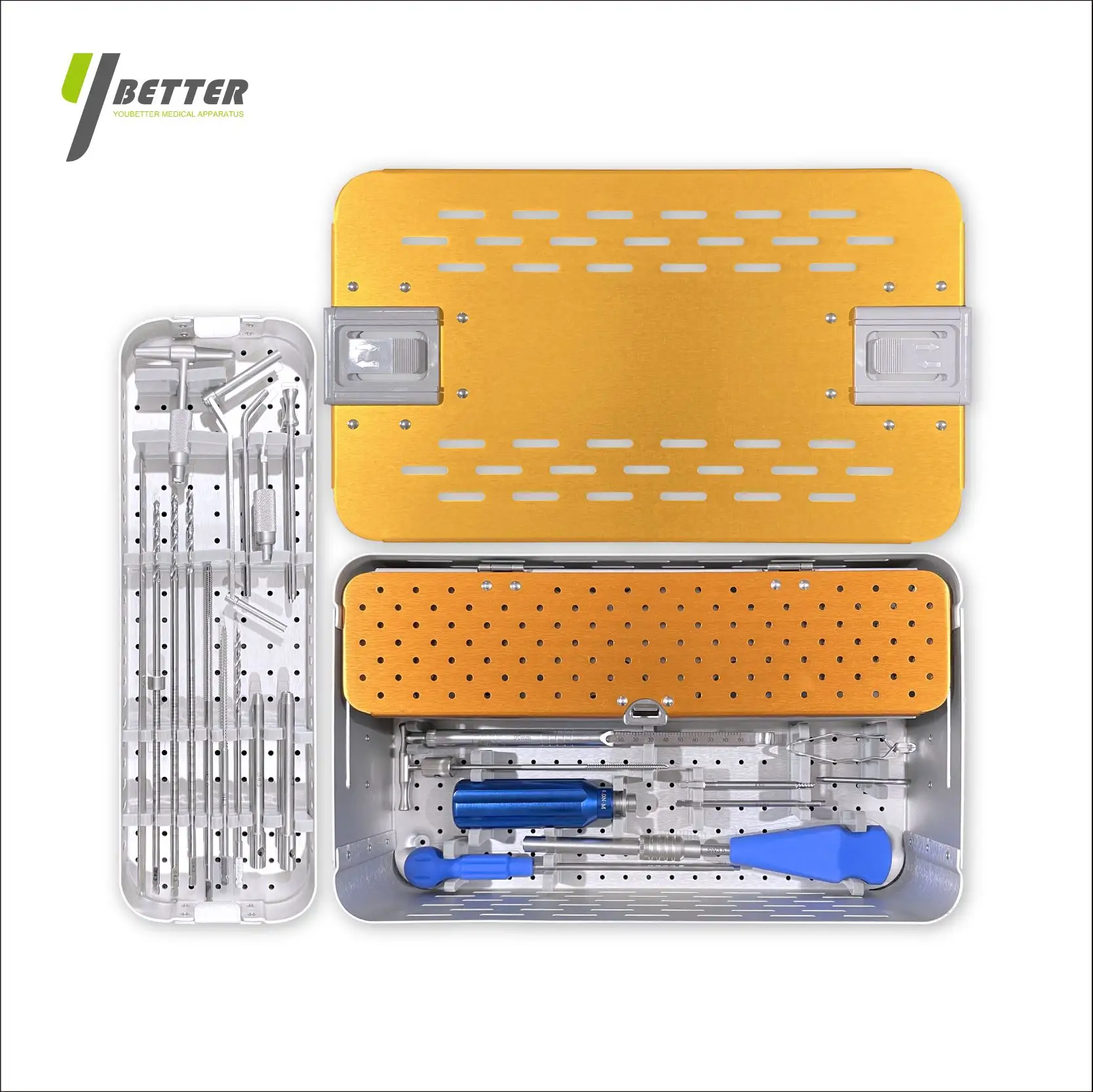
Scoliosis correction surgery is one of the most technically demanding procedures in modern spinal orthopedics. The ability to realign a severely curved spine, restore sagittal balance, and maintain long-term structural integrity depends heavily on the instrumentation used. Among all the hardware involved, the pedicle screw system plays the most central role in achieving and sustaining the desired correction. Understanding how these implants function within a scoliosis correction procedure provides essential insight for surgeons, procurement specialists, and clinical decision-makers evaluating spinal fixation solutions.
The pedicle screw system has transformed spinal deformity surgery since its widespread adoption. By anchoring directly into the pedicle — the strongest bony corridor of the vertebra — these systems offer three-dimensional control over each instrumented segment. This biomechanical advantage is what makes the pedicle screw system the preferred fixation strategy in nearly all modern scoliosis correction surgeries. This article explains the mechanism, clinical logic, surgical workflow, and technical considerations that make this instrumentation indispensable in deformity correction.
The Biomechanical Foundation of a Pedicle Screw System in Deformity Correction
Three-Dimensional Control Over Spinal Segments
What distinguishes a pedicle screw system from earlier hook-and-rod or wire-based constructs is its capacity to control the vertebra in all three planes simultaneously. Earlier instrumentation systems could apply corrective forces in only one or two planes, which limited the degree of correction achievable, particularly in thoracic scoliosis with significant rotational deformity. The pedicle screw, by engaging all three columns of the spine, enables surgeons to apply distraction, compression, translation, and derotation forces with far greater precision.
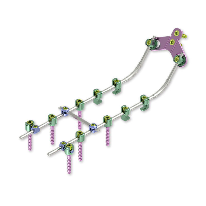
In scoliosis, the deformity is not simply a lateral curve — it involves vertebral rotation and loss of normal sagittal contour as well. The pedicle screw system addresses all of these components. When screws are placed bilaterally across multiple levels, they create a rigid foundation from which the connecting rods can be rotated, contoured, and tensioned to progressively straighten and derotate the spine. This triplanar control is the biomechanical reason why pedicle-based constructs consistently achieve superior correction rates compared to earlier techniques.
Load Distribution and Structural Stability
One of the critical mechanical functions of a pedicle screw system is how it distributes the corrective and maintenance loads across the construct. In scoliosis surgery, very significant forces are applied to the spine during correction maneuvers, and the instrumentation must withstand these forces without failure at the bone-implant interface. Because the pedicle is dense cortical bone with excellent purchase potential, screws placed through this corridor carry substantially higher pullout resistance than hooks or sublaminar wires.
This superior pullout strength allows the surgeon to apply more aggressive correction maneuvers during the operation and to rely on the construct for long-term maintenance of correction during the fusion healing period. The pedicle screw system transfers loads evenly along the rod-screw interface across multiple levels, reducing the peak stress at any single point. Well-designed locking mechanisms at the screw-rod junction ensure that the achieved correction is rigidly locked in place once the optimal alignment is confirmed.
How the Pedicle Screw System Supports Surgical Correction Techniques
Rod Rotation and In Situ Contouring
The surgical technique most associated with pedicle screw system constructs in scoliosis is the rod rotation maneuver. In this technique, the surgeon pre-contours a rod to match the desired postoperative sagittal profile and connects it to the screw heads on the concave side of the curve. By rotating this pre-bent rod from the coronal to the sagittal plane, the surgeon converts the lateral curve into the correct sagittal alignment, simultaneously reducing the Cobb angle and restoring normal thoracic kyphosis or lumbar lordosis.
This maneuver is only possible because the pedicle screw system allows the rod to be securely engaged with polyaxial or monoaxial screw heads that can be locked after the rotation is complete. The rigid engagement of every pedicle screw in the construct ensures that the rotational correction translates uniformly across each instrumented vertebra. Any looseness or inadequate purchase at a single screw level can compromise the entire correction, which is why implant quality and accurate screw placement are both critical to the success of this technique.
Derotation Maneuvers and Apical Correction
In more severe or rigid curves, direct vertebral derotation is an additional technique made possible by the pedicle screw system. With screws placed on both the concave and convex sides at the apical vertebra and adjacent levels, the surgeon can use derotation instruments attached to the screw extenders to rotate individual vertebrae back toward their neutral position. This directly addresses the rotational component of scoliosis that is responsible for the cosmetically significant rib hump and thoracic asymmetry.
Apical correction maneuvers demonstrate one of the most important surgical advantages of a comprehensive pedicle screw system: the ability to apply corrective forces at any specific level within the construct independently of other levels. This segmental control gives experienced surgeons the flexibility to achieve a more balanced, natural correction across the entire instrumented segment. The result is not only a better Cobb angle on imaging but also improved rotational correction and a more symmetrical clinical appearance for the patient.
Instrumentation Selection and Construct Planning for Scoliosis
Screw Diameter, Length, and Material Considerations
Selecting the appropriate pedicle screw system components for a scoliosis case requires careful preoperative planning based on imaging measurements and the patient's bone anatomy. Pedicle dimensions vary considerably across the thoracic and lumbar spine, and in pediatric patients with scoliosis, pedicles may be significantly narrowed or dysplastic at the apical levels. Computed tomography measurement of pedicle width, height, and angulation is essential to select screw diameters and trajectories that maximize purchase while avoiding cortical breach.
Rod diameter and material selection are equally important in the overall pedicle screw system construct. Stiffer rods produce larger corrective forces but require more strength for intraoperative bending and contouring. Softer alloys may reduce correction force transmission but are easier to handle. The choice between titanium alloy and cobalt-chromium rods involves trade-offs in stiffness, MRI compatibility, and fatigue resistance that the surgical team must evaluate based on the specific deformity and the planned fusion length.
Fusion Level Selection and Construct Length
The fusion level selection in scoliosis surgery directly determines how many segments will be instrumented with the pedicle screw system. Including too few levels risks adding-on deformity or proximal junctional failure, while extending the fusion unnecessarily sacrifices spinal mobility and functional range of motion. Established criteria guide surgeons in selecting stable end vertebrae, neutral vertebrae, and the lowest instrumented vertebra based on the flexibility and structural end-point of the curve.
Once the fusion levels are defined, the density of pedicle screw system placement across the construct affects both the strength of correction achievable and the overall construct rigidity. Higher screw density — placing screws at every instrumented level bilaterally — maximizes correction potential and reduces the risk of rod fracture by distributing stress across more anchor points. Selective screw placement strategies can reduce operative time and implant cost, but the decision must account for the stiffness of the curve and the planned correction technique.
Clinical Outcomes and Long-Term Reliability of Pedicle Screw Constructs
Correction Rate and Fusion Success
The clinical evidence supporting the pedicle screw system in scoliosis surgery is extensive. Studies across adolescent idiopathic scoliosis, adult degenerative scoliosis, and neuromuscular scoliosis consistently report superior correction rates with pedicle screw-based constructs compared to hybrid or hook-based systems. Correction of 60 to 70 percent of the primary curve Cobb angle is commonly achievable with all-screw constructs in flexible adolescent curves, with lower but clinically significant corrections in rigid adult deformities.
Long-term fusion outcomes are closely tied to the stability of the pedicle screw system construct during the healing period. Rigid fixation minimizes micromotion at the fusion interfaces, allowing consistent bone graft incorporation and solid arthrodesis across all instrumented levels. Once fusion is complete, the construct continues to maintain alignment against any residual deforming forces. Loss of correction over time is generally associated with pseudarthrosis or hardware failure, both of which are significantly less common with well-designed pedicle screw constructs.
Hardware Reliability and Complication Management
While the pedicle screw system delivers consistently strong outcomes, implant-related complications can occur and must be managed with experience and appropriate hardware selection. Rod fracture remains one of the more common late complications, particularly in adult deformity cases with long fusions and significant sagittal imbalance correction. The use of larger-diameter rods, satellite rod augmentation at the lumbosacral junction, and adequate screw density significantly reduces the risk of rod fracture in mechanically demanding constructs.
Screw loosening or pullout, while uncommon in patients with normal bone density, is a more significant concern in elderly patients with osteoporosis or in patients with neuromuscular scoliosis who have poor bone quality. Strategies such as cement augmentation, larger-diameter screws, and expanded surface area designs within the pedicle screw system can compensate for reduced bone quality and maintain adequate fixation strength. The ongoing evolution of implant design continues to address these challenges and extend the reliable application of pedicle screw constructs to a broader patient population.
Preoperative Planning and Intraoperative Navigation in Pedicle Screw Placement
Imaging-Based Planning for Accurate Screw Trajectories
Accurate preoperative planning is foundational to successful pedicle screw system placement in scoliosis surgery. In a rotated, curved spine, the pedicle axes deviate significantly from standard anatomical landmarks, making freehand screw placement based on surface anatomy alone unreliable in many cases. Preoperative CT-based planning software allows the surgical team to define the optimal entry point, trajectory, diameter, and length for each screw across all planned levels before the patient enters the operating room.
This detailed planning also helps identify levels where pedicle anatomy may preclude safe screw placement, allowing the surgeon to pre-plan alternative anchor strategies such as transverse process hooks or pedicle hooks at those specific segments. Incorporating these decisions into the preoperative plan ensures a coherent pedicle screw system construct design that accounts for anatomical variation without compromising the overall correction strategy.
Intraoperative Navigation and Robotic Assistance
Intraoperative navigation systems have significantly enhanced the accuracy and safety of pedicle screw system placement in scoliosis surgery. Fluoroscopy-based guidance, CT-based navigation, and robot-assisted platforms all provide real-time confirmation of screw trajectory before and during insertion. Studies consistently show reduced rates of cortical breach with navigated placement compared to freehand technique, particularly at the apical and thoracic levels where pedicle dimensions are smallest and rotational deformity is greatest.
Beyond accuracy, navigation reduces the radiation exposure experienced by the surgical team during complex multilevel procedures. As robotic platforms become more widely accessible, the ability to preplan and execute pedicle screw trajectories with submillimeter precision is increasingly available even for less experienced centers. The combination of rigorous preoperative planning and intraoperative navigation represents the current standard of excellence in pedicle screw system application for scoliosis surgery and continues to push clinical outcomes further.
FAQ
What makes a pedicle screw system more effective than older hook-based constructs for scoliosis?
A pedicle screw system provides three-dimensional control over each vertebra by engaging all three spinal columns simultaneously. This allows surgeons to apply corrective forces in all planes — lateral, axial, and rotational — which is not achievable with hook-based systems that anchor only to posterior elements. The result is consistently superior correction rates and more reliable long-term maintenance of alignment.
How does the pedicle screw system help correct the rotational component of scoliosis?
The rotational deformity in scoliosis is addressed through direct vertebral derotation maneuvers that are only possible when screws are placed bilaterally at the apical and adjacent levels. By attaching derotation instruments to the screw extenders, the surgeon can rotate individual vertebrae back toward neutral position. This technique is a core advantage of the pedicle screw system and directly reduces the rib hump and thoracic asymmetry associated with scoliosis.
Are pedicle screw systems safe for use in pediatric scoliosis patients?
Yes, pedicle screw system constructs are routinely used in adolescent idiopathic scoliosis and are supported by extensive safety data in pediatric populations. Careful preoperative CT planning and intraoperative navigation reduce the risk of cortical breach even in smaller pediatric pedicles. The implants are available in sizes appropriate for pediatric anatomy, and long-term follow-up studies confirm the durability and reliability of pedicle screw fixation in growing and skeletally mature adolescent patients.
How long does the pedicle screw system remain in the body after scoliosis surgery?
In most cases, the pedicle screw system remains permanently in the body after scoliosis surgery. Once solid fusion is achieved — typically within 12 to 18 months — the instrumentation is no longer mechanically necessary but is generally left in place unless it causes symptoms such as hardware prominence or pain. Removal is considered only when there is a specific clinical indication, as the risks of a second operation typically outweigh the benefits of routine hardware removal in asymptomatic patients.
Table of Contents
- The Biomechanical Foundation of a Pedicle Screw System in Deformity Correction
- How the Pedicle Screw System Supports Surgical Correction Techniques
- Instrumentation Selection and Construct Planning for Scoliosis
- Clinical Outcomes and Long-Term Reliability of Pedicle Screw Constructs
- Preoperative Planning and Intraoperative Navigation in Pedicle Screw Placement
-
FAQ
- What makes a pedicle screw system more effective than older hook-based constructs for scoliosis?
- How does the pedicle screw system help correct the rotational component of scoliosis?
- Are pedicle screw systems safe for use in pediatric scoliosis patients?
- How long does the pedicle screw system remain in the body after scoliosis surgery?