
 EN
EN
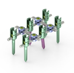
When a surgeon places a spinal screw into osteoporotic bone, the mechanical environment is fundamentally different from that of healthy vertebral bone. Osteoporosis reduces bone mineral density and disrupts trabecular architecture, leaving the cancellous core of a vertebral body with far less material to grip a spinal screw. The result is a measurable drop in pullout strength, toggling resistance, and long-term fixation reliability. Understanding exactly how a spinal screw behaves under these conditions is essential for any clinical team managing degenerative or traumatic spinal pathology in an aging population.
The clinical stakes are high. A failed spinal screw in an osteoporotic patient can lead to construct loosening, adjacent segment overload, or the need for painful revision surgery. Yet the procedure is frequently unavoidable because osteoporotic patients often suffer the very vertebral fractures and instabilities that require surgical stabilization. This article examines the performance profile of a spinal screw in low-density bone, the biological and mechanical reasons for fixation loss, and the augmentation strategies that modern spine surgery uses to restore reliable screw purchase in compromised vertebrae.
Why Osteoporosis Undermines Spinal Screw Fixation
The Mechanical Basis of Reduced Purchase
A spinal screw achieves fixation primarily through the interlocking of its threads with surrounding trabecular bone. In healthy vertebrae, the dense cancellous network distributes stress broadly around the spinal screw shaft. In osteoporotic bone, the same cancellous network is thinned and perforated, so the contact area between the spinal screw thread and bone is sharply reduced. Biomechanical studies consistently show that pullout force for a standard spinal screw drops by 30 to 60 percent when bone mineral density falls below the osteoporotic threshold. Toggling under cyclic loading accelerates this degradation, widening the interface void around the spinal screw and triggering micro-motion that prevents biological integration.
Cortical Purchase and Bicortical Techniques
One compensatory strategy is to advance the spinal screw tip through the far cortex of the vertebral body, achieving bicortical purchase. Cortical bone retains significantly more strength than cancellous bone even in osteoporotic patients, so engaging both cortices with the spinal screw tip can substantially improve axial pullout resistance. The tradeoff is an elevated risk of anterior vascular injury, particularly in the lumbar spine. Surgeons must therefore weigh the incremental mechanical gain of bicortical spinal screw placement against the anatomical hazards specific to each vertebral level. In the thoracic spine, where the spinal cord lies immediately behind the posterior cortex, the margin for error is even narrower.
Bone Cement Augmentation for Spinal Screw Stability
How Cement Augmentation Changes the Fixation Environment
Polymethylmethacrylate cement augmentation has become the most widely adopted method for strengthening a spinal screw in osteoporotic bone. In this technique, a cannulated spinal screw is inserted through a fenestrated shaft, and low-viscosity cement is injected under controlled pressure through the spinal screw itself. The cement fills the trabecular voids surrounding the spinal screw and polymerizes into a rigid mantle that dramatically increases the effective contact area. Pullout strength of an augmented spinal screw has been reported to match or exceed that of a conventional spinal screw in normal-density bone. This makes cement-augmented fixation a reliable option when osteoporosis would otherwise render the construct inadequate.
Clinical Considerations for Cement-Augmented Spinal Screw Use
The volume and viscosity of cement injected through the spinal screw must be carefully controlled. Excessive cement volume raises the risk of epidural or vascular leakage, which can cause neurological injury or pulmonary embolism. Fluoroscopic or CT guidance during spinal screw augmentation helps the surgeon monitor cement flow in real time and halt injection before extravasation occurs. Fenestrated spinal screw designs with multiple small side ports, rather than a single distal aperture, promote more uniform cement distribution around the spinal screw shaft and reduce the chance of directed leakage toward the spinal canal. Patient selection also matters: a spinal screw augmentation procedure carries greater cement leakage risk when the posterior vertebral wall is fractured or deficient.
Advanced Spinal Screw Designs for Osteoporotic Bone
Expandable and Variable-Angle Screw Designs
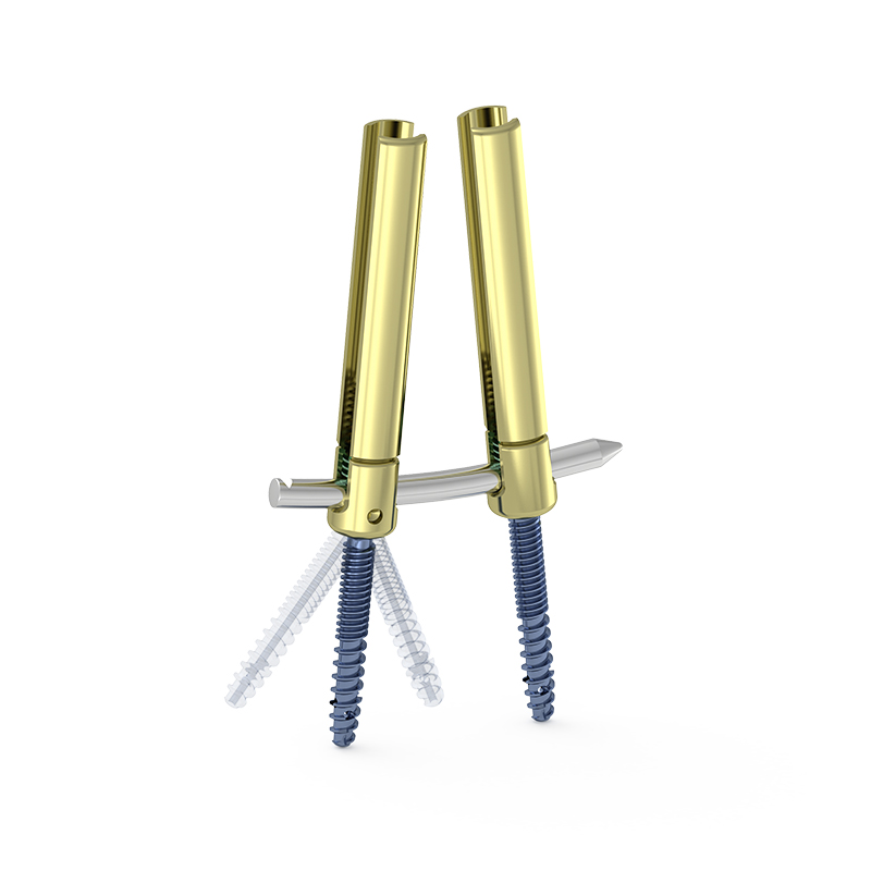
Beyond cement augmentation, implant engineers have developed spinal screw geometries specifically intended to perform better in low-density bone. Expandable spinal screw systems deploy radial wings or blades after insertion, mechanically interlocking with the surrounding trabecular structure in a way that a conventional spinal screw cannot replicate. This expansion distributes the load over a larger bone volume, reducing stress concentration at the thread-bone interface. Variable-angle spinal screw systems allow the surgeon to optimize trajectory after initial placement, which is valuable when the planned entry point offers suboptimal bone density and a slightly altered path would engage denser local bone. Both design families aim to make the spinal screw more tolerant of the irregular density distribution seen in osteoporotic vertebrae.
Cortical Bone Trajectory Techniques
The cortical bone trajectory technique repositions the spinal screw path so that the screw travels through a greater length of dense cortical bone within the pedicle and posterior vertebral body. A standard pedicle spinal screw follows a medially directed trajectory through primarily cancellous tissue. The cortical trajectory spinal screw starts more laterally and angles steeply medial and cephalad, maximizing engagement with the dense cortical pedicle walls. Studies in osteoporotic cadaveric specimens show that a cortical trajectory spinal screw can achieve pullout strength comparable to a standard pedicle spinal screw in normal bone, even when the host vertebra is severely osteoporotic. This makes the technique particularly attractive for minimally invasive lumbar fusion in elderly patients.
FAQ
Why does a standard spinal screw loosen more quickly in osteoporotic patients?
Osteoporotic bone has reduced trabecular density and weaker cortical shells, giving the spinal screw less material to grip. Under repetitive loading, the reduced bone-screw interface allows micro-motion that progressively enlarges the cavity around the spinal screw, accelerating loosening compared to fixation in normal-density bone.
Is bone cement augmentation safe for every patient requiring a spinal screw in osteoporotic bone?
Cement augmentation improves spinal screw pullout strength significantly, but it is not universally appropriate. Patients with a disrupted posterior vertebral wall, known allergy to cement components, or severe cardiopulmonary compromise require individualized risk assessment before a cement-augmented spinal screw procedure is planned. Imaging-guided injection and careful volume control remain essential safety measures.
How do surgeons decide which spinal screw technique to use in an osteoporotic patient?
The decision typically weighs preoperative bone mineral density measurement, the spinal level being instrumented, the degree of deformity correction needed, and the patient's overall health status. A severely osteoporotic patient undergoing multi-level lumbar fusion may benefit from both cement augmentation and a cortical trajectory spinal screw approach, while a mildly osteoporotic patient undergoing a single-level procedure may be managed with an expandable spinal screw design alone.