
 EN
EN
Selecting the correct pedicle screw size for lumbar fusion procedures is one of the most consequential decisions a spine surgeon makes during operative planning. The diameter, length, and thread design of each pedicle screw directly influence fixation strength, load distribution across the construct, and ultimately the patient's long-term clinical outcome. A mismatch between implant dimensions and patient anatomy does not merely reduce mechanical performance — it can lead to cortical breach, nerve root injury, implant loosening, or the need for costly revision surgery.
Understanding how to size a pedicle screw correctly requires integrating preoperative imaging data, biomechanical principles, and an accurate understanding of lumbar vertebral anatomy across different levels. This guide walks through the critical sizing factors surgeons and procurement specialists should consider, from interpreting CT measurements to selecting screw geometry for specific fusion indications. Whether you are planning a single-level L4-L5 fusion or a multi-segment reconstruction, the logic for choosing each pedicle screw remains grounded in the same core methodology.
Understanding Lumbar Pedicle Anatomy and Its Role in Screw Sizing
Regional Variation Across Lumbar Levels
The lumbar spine is not a uniform structure, and pedicle screw sizing must account for the significant anatomical variation across L1 through L5. The pedicle isthmus — the narrowest cross-sectional area through which the screw must safely pass — progressively widens from the upper lumbar vertebrae to the lower levels. At L5, pedicle widths can approach 18 to 20 millimeters in some patients, while L1 pedicles may measure as narrow as 7 to 9 millimeters in smaller individuals.
This regional variation means that a single screw diameter selected for the entire lumbar construct is rarely appropriate. Surgeons must plan each level independently, using CT axial cuts to measure the true pedicle transverse diameter and determine safe screw corridor dimensions. Relying on generic size charts without patient-specific imaging significantly increases the risk of medial or lateral cortical breach during pedicle screw placement.
Vertebral body depth also varies across lumbar levels and influences the optimal screw length. A pedicle screw that engages the anterior cortex of the vertebral body achieves three-point fixation and substantially greater pullout resistance than one that stops short of the anterior third of the body. Accurate measurement of the pedicle-to-anterior-cortex distance on sagittal CT reconstructions is therefore essential for length selection.
Bone Density Considerations in Screw Diameter Selection
Bone mineral density plays a decisive role in determining the appropriate pedicle screw diameter for any given patient. In patients with normal bone quality, a standard diameter that fills approximately 80 percent of the pedicle isthmus provides reliable cortical purchase without undue risk of fracture. However, in patients with osteopenia or osteoporosis — populations increasingly common in lumbar fusion practice — standard diameter screws may not generate adequate pullout force in trabecular bone alone.
For osteoporotic patients, upsizing the pedicle screw diameter to maximize cortical engagement, using fenestrated screws with cement augmentation, or selecting screws with expanded thread designs can compensate for reduced bone quality. These decisions should be based on preoperative DEXA scan findings and intraoperative tactile feedback during pedicle preparation. Ignoring bone density when selecting screw dimensions is a common source of early construct failure in the elderly lumbar fusion patient.
Key Dimensional Parameters: Diameter and Length
Choosing the Correct Screw Diameter
The outer diameter of the pedicle screw is the single most important dimension in determining fixation quality. Standard lumbar pedicle screws range from 5.5 millimeters to 8.5 millimeters in outer diameter, with 6.5 and 7.0 millimeter options being among the most commonly deployed in adult lumbar fusion. The diameter should ideally correspond to approximately 80 to 85 percent of the measured pedicle transverse width to achieve cortical apposition without breaching the pedicle wall.
Surgeons frequently choose between standard and cannulated pedicle screw designs, particularly when fluoroscopic or navigation-guided placement is planned. Cannulated screws allow guidewire-assisted insertion and offer added safety in challenging anatomies or revision cases where prior instrumentation has altered the native pedicle corridor. The choice of cannulated versus solid core design does not typically alter the diameter selection logic but should be factored into overall screw strength calculations.
In practice, it is advisable to have both the planned diameter and one size larger available on the sterile field before incision. Intraoperative findings — including unexpected screw toggling or feel of inadequate thread engagement — may necessitate upsizing to achieve satisfactory fixation. Having contingency sizing prepared avoids delays and ensures the surgeon can respond to real-time anatomical information during pedicle screw placement.
Determining Appropriate Screw Length
Screw length for lumbar fusion is typically selected to achieve bicortical or near-bicortical purchase, meaning the tip of the pedicle screw reaches the anterior one-third of the vertebral body without penetrating the anterior cortex. Standard lumbar pedicle screw lengths range from 35 millimeters to 60 millimeters, with 40 to 50 millimeter lengths being most common in routine adult lumbar fusion at L3 through L5.
Pedicle length measurements should be taken from sagittal CT reconstructions with the measurement line following the planned screw trajectory rather than parallel to the endplate. An oblique trajectory, particularly in the axial plane when aiming medially for anatomical reasons, effectively increases the true screw path compared to a straight anteroposterior measurement. Failure to account for trajectory angulation frequently results in selecting a pedicle screw that is 5 to 10 millimeters shorter than optimal, reducing the purchase achieved in the anterior vertebral body cortex.
At L5, longer screws are more often feasible due to the wider and deeper vertebral body, while at upper lumbar levels the available depth may be more restricted. In all cases, the goal is to maximize the length of screw-bone contact within the safe corridor. A pedicle screw that is correctly sized for length but too short in diameter will still demonstrate inferior pullout performance, underscoring that both dimensions must be optimized simultaneously.
The Role of Screw Design and Thread Geometry
Thread Pitch and Pullout Resistance
Beyond outer diameter and length, the thread design of a pedicle screw is a critical determinant of biomechanical performance. Thread pitch — the distance between adjacent thread crests — affects how efficiently the screw converts rotational insertion torque into axial clamping force. Fine-pitch threads engage more bone per unit of screw length and generally produce higher pullout resistance in cortical bone, while coarser threads are more effective in cancellous bone because they displace and compact a greater volume of trabecular material.
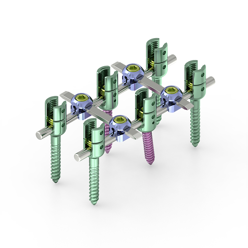
Most modern lumbar pedicle screw systems use a dual-lead or variable-pitch thread design that attempts to optimize performance across both cortical and cancellous bone zones encountered along the pedicle and vertebral body. These hybrid thread geometries have shown improved resistance to cyclic loading in biomechanical testing, which is particularly relevant in multi-level lumbar fusion constructs where each screw experiences complex combined loading from axial compression, bending, and torsion during patient activity.
Head Design and Reduction Capability
The head design of the pedicle screw is equally important in lumbar fusion, especially when deformity correction, spondylolisthesis reduction, or multi-level alignment restoration is part of the surgical objective. Polyaxial screw heads allow the rod to be captured across a range of angulations without requiring precise rod bending, simplifying rod insertion and reducing operative time in complex constructs. However, polyaxial heads introduce a degree of rotational freedom that can complicate correction maneuvers.
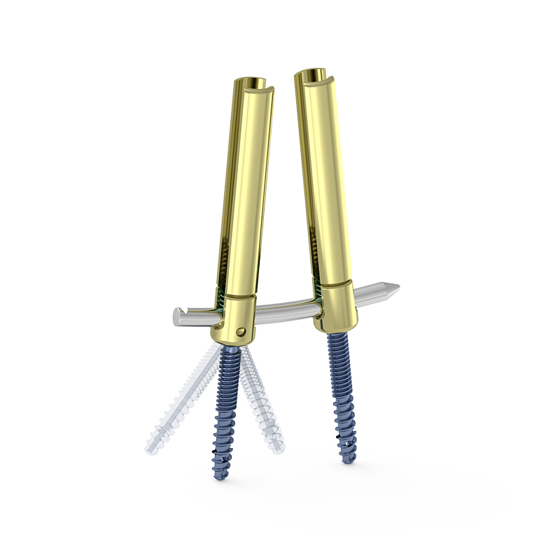
For cases involving spondylolisthesis or significant sagittal imbalance, reduction-type pedicle screw designs provide a distinct mechanical advantage. A pedicle screw with a tall reduction tower allows the surgeon to translate the slipped vertebra into alignment with the adjacent level by sequentially tightening the head down toward the rod, generating controlled corrective force without requiring significant manual vertebral manipulation. Choosing a reduction design when the anatomy demands it is not merely a convenience — it is an important safety consideration that reduces the risk of iatrogenic neurological injury during reduction maneuvers.
The choice between standard, reduction, and uniplanar head designs should be made during preoperative planning based on the degree of deformity, the levels involved, and the planned correction strategy. A surgeon who defaults to the same head design for every lumbar fusion case regardless of pathology is underutilizing the biomechanical tools available in contemporary pedicle screw systems.
Preoperative Planning Workflow for Accurate Sizing
Imaging-Based Measurement Protocols
A reliable pedicle screw sizing workflow begins with high-quality preoperative CT imaging. Axial slices at each planned fusion level should be reviewed to measure the narrowest pedicle transverse width and the medial-to-lateral pedicle angulation. These measurements establish the maximum safe outer diameter and guide the planned insertion trajectory. Sagittal reconstructions are used to measure pedicle-to-anterior-cortex depth along the intended screw axis and to identify any deformity, fractured endplates, or prior hardware that may alter the available corridor.
Surgeons increasingly use spinal navigation platforms and robotic assistance to execute preoperatively planned pedicle screw trajectories with high fidelity. These technologies allow the planned diameter and length from the CT-based planning stage to be confirmed intraoperatively and reduce the incidence of cortical breaches compared to freehand techniques. Even when advanced navigation is not available, meticulous CT measurement remains the foundation of every sound sizing decision.
Intraoperative Verification and Adjustment
Even with thorough preoperative planning, intraoperative verification remains a non-negotiable step before final pedicle screw seating. Fluoroscopic confirmation in both anteroposterior and lateral planes after pedicle preparation allows the surgeon to verify trajectory before committing to a screw diameter. Medial wall probing of the pedicle channel provides tactile feedback about corridor integrity and can reveal a breach before any screw is placed.
If the prepared channel feels loose or the planned diameter appears small relative to the actual anatomy encountered, upsizing by 0.5 to 1 millimeter in diameter is a reasonable intraoperative adjustment. Conversely, if the pedicle appears smaller than preoperative imaging suggested — a situation that can occur with rotational deformity or imaging artifact — downsizing to avoid cortical perforation is the appropriate response. The flexibility to modify pedicle screw size intraoperatively is only possible when the instrument set includes a full range of sizes on the sterile field.
Electromyographic monitoring during pedicle preparation and screw insertion adds another layer of safety by detecting proximity to nerve roots before injury occurs. In revision lumbar fusion cases where anatomy is distorted and pedicle corridors may be partially filled with fibrous tissue or prior implant tracks, this monitoring is particularly valuable and should influence the final pedicle screw size and position decisions.
Special Sizing Considerations in Complex Lumbar Fusion Cases
Revision Surgery and Altered Pedicle Anatomy
Revision lumbar fusion presents unique challenges for pedicle screw sizing because prior instrumentation may have partially remodeled or weakened the pedicle bone. A previously placed screw leaves behind a void that may not fully remodel with bone, meaning a same-size screw in the same position will achieve substantially inferior fixation. In revision cases, surgeons often upsize the pedicle screw diameter by at least 1 millimeter to engage fresh cortical bone outside the prior track.
Alternatively, supplemental techniques such as off-axis pedicle screw placement, accessory cortical trajectory screws at adjacent vertebral levels, or vertebral body augmentation may be required when upsizing alone cannot restore adequate fixation. The key principle is that revision sizing is never simply a repetition of the primary case — it requires a fresh anatomical assessment and often a fundamentally different sizing strategy to achieve a stable pedicle screw construct.
Pediatric and Smaller Adult Anatomies
Pediatric spine surgery and procedures in smaller adult patients present the opposite sizing challenge: pedicles that are narrow enough that even the smallest standard pedicle screw diameter risks medial or lateral wall perforation. In these cases, CT-based morphometric analysis is even more critical, and surgeons may need to select undersized screws, use in-situ bending techniques, or consider alternative fixation strategies such as sublaminar bands or hooks at certain levels.
Mini-open and minimally invasive lumbar fusion approaches do not change the fundamental sizing logic but do restrict intraoperative visualization, making preoperative planning accuracy even more important. When working through a tubular retractor system, the surgeon has less ability to visually confirm trajectory and must rely more heavily on image guidance and preoperatively determined pedicle screw dimensions to execute placement safely.
FAQ
What is the most important factor when selecting pedicle screw diameter for lumbar fusion?
The most critical factor is the measured transverse width of the pedicle isthmus at each specific lumbar level, obtained from preoperative CT imaging. The selected pedicle screw outer diameter should fill approximately 80 to 85 percent of this measured width to achieve cortical apposition without risking medial or lateral wall breach. Bone density is the second most important variable, as it influences the adequacy of thread purchase within the available screw corridor.
How do I determine the correct pedicle screw length for lumbar fusion?
Screw length should be selected to allow the tip of the pedicle screw to reach the anterior one-third of the vertebral body, measured along the planned insertion trajectory on sagittal CT reconstructions. This achieves near-bicortical purchase and maximizes pullout resistance. It is important to measure along the actual screw path rather than in a straight anteroposterior direction, as trajectory angulation effectively increases the required screw length by 5 to 10 millimeters in many cases.
When should a reduction-type pedicle screw be chosen over a standard design?
A reduction-type pedicle screw is indicated when the surgical plan includes correction of spondylolisthesis, significant sagittal imbalance, or vertebral translation at one or more fusion levels. The extended reduction tower allows the surgeon to gradually bring the displaced vertebra into alignment with the rod without applying direct manual force to the neural elements, reducing the risk of neurological injury during the correction maneuver. This design choice should be made during preoperative planning, not improvised intraoperatively.
Can pedicle screw size affect fusion outcomes over the long term?
Yes, undersized or oversized pedicle screw selection can directly affect long-term fusion outcomes. An undersized screw may toggle under cyclical loading and promote fibrous encapsulation rather than solid osseointegration, potentially leading to pseudarthrosis. An oversized screw that breaches the pedicle wall can cause chronic nerve root irritation or compromise the structural integrity of the pedicle itself. Accurate sizing that matches the patient's anatomy and bone quality is therefore directly linked to achieving durable fusion and a favorable clinical result.
Table of Contents
- Understanding Lumbar Pedicle Anatomy and Its Role in Screw Sizing
- Key Dimensional Parameters: Diameter and Length
- The Role of Screw Design and Thread Geometry
- Preoperative Planning Workflow for Accurate Sizing
- Special Sizing Considerations in Complex Lumbar Fusion Cases
-
FAQ
- What is the most important factor when selecting pedicle screw diameter for lumbar fusion?
- How do I determine the correct pedicle screw length for lumbar fusion?
- When should a reduction-type pedicle screw be chosen over a standard design?
- Can pedicle screw size affect fusion outcomes over the long term?