
 EN
EN
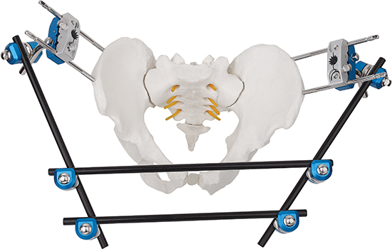
An external fixator is a mechanical device that plays a critical role in orthopedic trauma care by providing temporary stabilization of fractures and complex injuries. Unlike internal fixation methods, an external fixator works from outside the body, using pins and rods to immobilize bone fragments while allowing access to surrounding soft tissues. Understanding how an external fixator delivers this stabilization is essential for healthcare professionals, trauma teams, and patients facing acute orthopedic injuries.
The temporary nature of an external fixator makes it uniquely valuable in emergency orthopedic settings. When patients arrive with severe fractures, extensive soft tissue damage, or polytrauma, an external fixator can be applied rapidly to stabilize the injury without requiring extensive surgical dissection. This rapid deployment capability directly supports patient safety during the critical early phase of trauma management.
How an External Fixator Stabilizes Fractured Bone
Core Stabilization Mechanism
An external fixator achieves stabilization through a rigid frame structure that transfers mechanical load away from the injured bone. The system consists of percutaneous pins or wires inserted into healthy bone proximal and distal to the fracture site. These pins connect to an external frame of bars and clamps, creating a bridging construct that prevents harmful motion at the fracture location. By maintaining precise alignment and minimizing micromotion, an external fixator allows the natural bone healing process to proceed in a biomechanically favorable environment.
The rigidity of an external fixator directly correlates with healing outcomes. When an external fixator frame maintains tight alignment and minimal deflection under load-bearing stress, callus formation progresses predictably. Conversely, excessive frame flexibility or pin loosening can delay healing and compromise outcomes. Modern external fixator designs incorporate stronger materials and advanced clamp systems to maximize this stabilization across various fracture configurations and patient weight categories.
Pin and Wire Placement Strategy
The success of an external fixator depends critically on optimal pin placement in strong cortical bone. Surgeons position pins in trajectories that avoid vital neurovascular structures, major vessels, nerves, and organs while ensuring adequate bone purchase. Each pin inserted into an external fixator must achieve bicortical purchase, meaning it penetrates both cortices of the bone, to maximize resistance to pull-out and shear forces. This strategic pin placement transforms an external fixator from a simple framework into a highly effective load-sharing device.
Pin spacing and the number of pins significantly impact external fixator performance. An external fixator typically requires at least two pins proximal and two pins distal to the fracture, though complex fractures may benefit from additional pins. Wider spacing between pins increases the mechanical advantage and reduces stress concentration at individual pin sites. When properly configured, an external fixator distributes forces evenly across multiple support points, reducing the risk of pin loosening and frame failure during the temporary stabilization period.
Clinical Applications and Temporary Use Cases
Trauma and Emergency Stabilization
In acute trauma settings, an external fixator serves as the gold standard for rapid temporary stabilization of unstable fractures. Polytrauma patients with multiple injuries, severe soft tissue damage, or hemodynamic instability benefit enormously from the speed of application that an external fixator provides. By quickly establishing fracture stability without prolonged anesthesia or extensive tissue handling, an external fixator allows trauma teams to shift focus to life-threatening injuries while maintaining fracture protection. This temporary approach significantly reduces complications such as fat embolism and infection during the critical first days post-injury.
High-energy mechanisms frequently produce complex fractures that require an external fixator as an interim solution. Pelvic fractures, femoral shaft fractures, and tibial plateau injuries respond exceptionally well to external fixator stabilization in emergency contexts. The temporary nature of external fixator support means that once the patient stabilizes and soft tissues recover, conversion to internal fixation can occur under optimal conditions with reduced infection and nonunion risk.
Soft Tissue Damage Management
When fractures accompany severe soft tissue trauma, burn injuries, or crush mechanisms, an external fixator provides unique advantages. Unlike internal fixation devices that require closure of compromised soft tissues, an external fixator keeps hardware external and accessible. This accessibility allows plastic surgeons and soft tissue specialists to work simultaneously on wound management, debridement, and reconstruction while an external fixator maintains fracture stability. The temporary nature of this arrangement prevents infection complications that might occur if metal implants were buried in damaged tissue.
Vascular injuries present another scenario where an external fixator excels. When fractures coexist with arterial damage requiring vascular repair, an external fixator stabilizes bone while vascular surgeons work without the constraint of internal fixation hardware. This parallel approach accelerates overall trauma management and improves limb salvage outcomes, demonstrating how external fixator temporary stabilization adapts to complex clinical scenarios.
Biomechanical Advantages and Load Management
Stiffness and Micromotion Control
The biomechanical superiority of an external fixator lies in its ability to control micromotion at the fracture site with precision. When bone fragments experience excessive motion, secondary fracture lines develop, delays in healing accelerate, and infection risk increases. An external fixator minimizes this micromotion through its rigid configuration, keeping relative displacement below the critical threshold where healing is impaired. Studies comparing various fixation methods demonstrate that an external fixator achieves micromotion levels compatible with optimal bone healing when properly applied.
Load sharing between the external fixator frame and the healing bone represents a key feature of temporary stabilization. As bone callus develops, stress gradually transfers from the external fixator to new osseous tissue, creating a natural transition toward weight-bearing. This progressive load transfer that an external fixator enables prevents stress shielding and promotes robust bone remodeling, resulting in mechanically competent healed bone. The temporary support window provided by an external fixator aligns perfectly with biological healing timelines, typically ranging from six to twelve weeks depending on fracture severity and patient factors.
Adjustability and Clinical Flexibility
A practical strength of an external fixator is its adjustability during the temporary stabilization phase. Unlike internal fixation locked immediately during surgery, an external fixator frame can be modified postoperatively to correct residual malalignment, compression, or rotation. Orthopedic surgeons can make fine adjustments using specialized clamp systems without additional anesthesia, improving alignment accuracy and reducing the need for revision procedures. This flexibility is particularly valuable when initial injury assessment is incomplete or when significant swelling affects anatomical landmarks during primary fixation.
Incremental distraction using an external fixator represents another advanced application for temporary stabilization. When fractures present with bone loss, comminution, or segmental defects, an external fixator can be progressively adjusted to achieve lengthening or compression docking of fragments. This temporary distraction capability allows soft tissue adaptation and promotes new bone formation through callotasis, a process uniquely enabled by external fixator devices. The ability to modulate this mechanical environment over weeks or months makes an external fixator invaluable for complex fracture management.
FAQ
How long can an external fixator remain in place for temporary stabilization?
An external fixator typically remains in place for six to sixteen weeks, depending on fracture type, location, and healing progression. For temporary stabilization in acute trauma, an external fixator may serve as a bridge for just one to three weeks until internal fixation conversion becomes feasible. In complex fractures managed by lengthening or compression techniques, an external fixator may function throughout the entire treatment course. Regular radiographic assessment guides decisions about when an external fixator can be safely removed or converted to other fixation methods.
What complications can occur with external fixator use during temporary stabilization?
Common complications include pin tract infections, pin loosening, and frame deformity if stability is inadequate. Vascular or nerve injury is rare but possible if an external fixator pins are placed in unsafe anatomical corridors. Stiffness in adjacent joints may develop if external fixator immobilization is prolonged, requiring aggressive physical therapy post-removal. Patient compliance with pin care and activity restrictions significantly influences whether complications develop during temporary external fixator treatment.
When should an external fixator be converted to internal fixation?
Conversion timing depends on fracture characteristics, soft tissue status, and clinical trajectory. External fixator conversion to internal fixation typically occurs once acute swelling resolves, skin quality improves, and medical stability allows extended surgery. For polytrauma patients, conversion may happen within two to four weeks after injury once life threats are resolved. For simple fractures managed temporarily, external fixator conversion might occur immediately or be deferred if the external fixator provides adequate definitive fixation. Individual patient assessment determines optimal conversion timing.