
 EN
EN
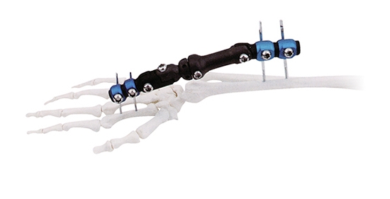
Soft tissue damage during fracture treatment remains one of the most significant concerns in orthopedic medicine. When bones break, the surrounding muscles, nerves, blood vessels, and connective tissue face additional trauma from traditional surgical interventions. An external fixator offers a scientifically proven approach to minimize this secondary injury while maintaining skeletal stability. Unlike internal fixation methods that require large incisions and direct manipulation of soft tissues, an external fixator works through a minimally invasive principle that keeps the surgical field external to the body.
The external fixator represents a fundamental shift in how orthopedic surgeons address complex fractures, particularly in polytrauma cases where multiple injuries coexist. By positioning fixation hardware outside the body and connecting it to the bone through percutaneous pins or wires, the external fixator eliminates the need for extensive soft tissue dissection. This preservation of soft tissue integrity directly translates to faster healing, reduced infection risk, and improved long-term functional outcomes for patients across diverse trauma scenarios.
Mechanical Principles Behind Soft Tissue Protection
Minimally Invasive Pin Placement
The external fixator achieves soft tissue protection through its fundamental design architecture. Percutaneous pins inserted through the external fixator bypass muscle layers and deep vascular structures by using precise trajectories established preoperatively. These pins require only small puncture wounds rather than the extensive surgical approaches needed for plate and screw fixation. Each external fixator configuration is planned to avoid neurovascular bundles, ensuring that the external fixator installation causes minimal collateral damage to vital structures surrounding the fracture site.
Surgeons utilizing the external fixator benefit from the ability to adjust pin trajectory in real time using fluoroscopic guidance. This precision means the external fixator can be positioned to navigate between muscle fascicles and around blood vessels without requiring wider exposure. The external fixator's external frame remains completely outside the surgical wound, allowing soft tissues to heal naturally without ongoing irritation from internal hardware.
Immediate Fracture Stability Without Dissection
Once the external fixator is installed, fracture fragments achieve immediate rigid stability through the rigid connecting frame. This stability is achieved without opening the fracture site itself, meaning the external fixator prevents the soft tissue damage that occurs when fracture edges are manipulated or when internal implants are inserted directly into bone. Soft tissues surrounding the fracture remain undisturbed, allowing their intrinsic healing mechanisms to function optimally while the external fixator maintains skeletal alignment.
The external fixator's load-sharing characteristics differ from rigid internal fixation. By allowing controlled micromotion at the fracture site, the external fixator stimulates callus formation and promotes biological healing. This external fixator principle reduces stress shielding, a phenomenon where internal implants can weaken bone remodeling, and instead facilitates natural healing processes while protecting soft tissues from excessive strain.
Clinical Advantages in Trauma and Complex Cases
Rapid Soft Tissue Recovery and Early Mobilization
Patients treated with an external fixator experience significantly faster soft tissue recovery compared to traditional open reduction and internal fixation approaches. Because the external fixator causes minimal tissue trauma, inflammatory response remains localized to only the pin sites rather than extending throughout the surgical field. This external fixator characteristic enables earlier functional rehabilitation, allowing physical therapists to mobilize adjacent joints while the external fixator maintains fracture stability.
Early mobilization supported by the external fixator framework reduces complications such as muscle atrophy, joint stiffness, and thromboembolism. The external fixator design allows patients to begin weight-bearing protocols sooner, which further accelerates soft tissue remodeling and restores functional capacity. In polytrauma patients where multiple injuries compete for healing resources, the external fixator's minimal tissue invasion preserves soft tissue viability for other injury sites.
Infection Prevention and Soft Tissue Health
Soft tissue infection represents one of the most serious complications in orthopedic trauma. The external fixator substantially reduces infection risk through two mechanisms: reduced surgical wound size and preservation of local blood supply. When the external fixator is applied, only small puncture wounds are created, limiting bacterial entry pathways compared to the extensive incisions required for plate fixation. The external fixator's percutaneous pin sites, while requiring hygiene protocols, present a dramatically smaller infection surface than deep surgical wounds.
The intact soft tissue envelope maintained by the external fixator preserves micro-vascular integrity and local immune function. This external fixator benefit becomes critical in contaminated or open fractures where tissue viability is already compromised. Surgeons can apply the external fixator rapidly in emergency settings, then reassess soft tissue status and plan delayed reconstruction without the additional damage that would result from removing internal fixation hardware.
Application Scenarios Where External Fixator Excels
Open Fractures and Contaminated Wounds
Open fractures with significant soft tissue stripping represent the quintessential indication for the external fixator approach. The external fixator provides immediate skeletal stability while leaving the traumatized soft tissue zone accessible for serial debridement and inspection. Surgeons can evaluate tissue viability repeatedly without disrupting fixation by using the external fixator framework as the stable anchor. This external fixator capability is impossible with internal fixation, which requires the fracture site to remain undisturbed once implants are inserted.
In high-energy trauma with massive soft tissue injury, the external fixator serves as a rapid, definitive fixation that buys time for tissue assessment and planning. The external fixator allows temporary bridging fixation in emergencies, with conversion to definitive fixation once the patient stabilizes and soft tissue status is better understood. This external fixator flexibility has transformed survival and functional outcomes in severe polytrauma cases.
Revision and Salvage Scenarios
When previous fixation attempts have failed or created significant soft tissue scarring, the external fixator offers a soft tissue-sparing alternative. The external fixator can be applied without requiring removal of failed internal hardware initially, allowing soft tissues to recover before staged revision. This external fixator strategy preserves tissue quality for future reconstructive procedures, whether osteogenic distraction or delayed arthroplasty.
Patients with chronic infections or non-unions complicated by soft tissue loss benefit from the external fixator's ability to maintain alignment while allowing aggressive soft tissue management. The external fixator keeps the operative field external, preventing hardware from becoming a nidus for persistent infection and enabling infection control without sacrificing stability.
FAQ
What is an external fixator and how does it work?
An external fixator is an orthopedic device consisting of a rigid metal frame positioned outside the body, connected to fractured bone through percutaneous pins or wires. The external fixator maintains skeletal alignment and stability without requiring internal implants. Surgeons insert pins into bone segments on either side of the fracture, then connect these pins to the external fixator frame through clamps and rods. This external fixator system immediately stabilizes the fracture while leaving soft tissues undisturbed.
How does external fixator prevent infection better than internal fixation?
The external fixator reduces infection through minimal surgical trauma and preserved soft tissue blood supply. Unlike internal fixation requiring large incisions that expose deep tissues, the external fixator uses only small puncture wounds for pin insertion. The external fixator preserves local micro-vascular networks that are critical for immune function and tissue healing. Pin site infections, though possible, are superficial and easily managed compared to deep surgical site infections associated with internal hardware.
Can external fixator be used for all fracture types?
While the external fixator is particularly valuable for open fractures, complex trauma, and soft tissue compromise, it is not ideal for all fracture patterns. Simple closed fractures without soft tissue injury may be better treated with internal fixation or conservative management. The external fixator excels in polytrauma, contaminated wounds, and revision cases where soft tissue preservation is paramount. Your surgeon will determine whether the external fixator is appropriate based on fracture characteristics, injury severity, and soft tissue status.