
 EN
EN
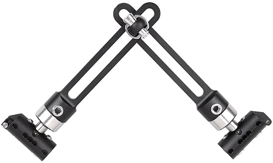
Limb lengthening represents one of the most complex orthopedic procedures in modern medicine, requiring precise control, gradual bone extension, and sustained stability throughout the healing process. The choice of fixation method directly influences surgical success, patient recovery time, and long-term functional outcomes. An external fixator has become the gold standard for this purpose, offering unparalleled advantages that internal fixation methods simply cannot match. Understanding why an external fixator is preferred reveals the critical balance between mechanical control, biological healing, and clinical practicality that defines contemporary limb lengthening surgery.
The external fixator provides a mechanical framework that allows orthopedic surgeons to control the rate and direction of bone extension with millimeter precision. Unlike internal hardware, which becomes buried within tissue and cannot be adjusted after initial placement, an external fixator remains accessible throughout the entire treatment duration. This accessibility transforms the clinical approach to limb lengthening from a one-time surgical event into a controlled, adjustable therapeutic process that can be modified based on radiographic findings, tissue response, and patient tolerance.
Biomechanical Precision and Distraction Control
Why External Fixators Enable Precise Bone Extension
The external fixator operates on a principle of gradual, controlled distraction that respects the biological limits of bone healing. When surgeons perform an osteotomy—a surgical bone cut—the external fixator maintains exact spacing between bone fragments through an adjustable frame system. This external fixator framework allows daily micro-adjustments, typically advancing the bone segments by one millimeter per day, a rate proven optimal for both bone formation and soft tissue adaptation. The precision that an external fixator offers prevents over-distraction, which would compromise bone healing, and under-distraction, which would fail to achieve the desired length.
Internal fixation devices, by contrast, establish a fixed gap that cannot be altered without reopening the surgical site. An external fixator solves this limitation entirely, making it the preferred choice for complex lengthening cases where the optimal distraction rate may need adjustment. The adjustable nature of an external fixator also accommodates individual biological variation—some patients' bones regenerate faster than others, and clinical adjustments to an external fixator accommodate these differences seamlessly without additional surgery.
Multi-Directional Control Capabilities
Modern external fixator systems provide hexapod designs that permit simultaneous control in six directions—three translational axes and three rotational axes. This multi-directional capability makes an external fixator invaluable when lengthening must occur while simultaneously correcting angular deformities, rotational malalignment, or existing anatomical distortions. Surgeons using an external fixator can progressively correct complex three-dimensional bone malalignment while extending limb length, a feat impossible with simpler fixation approaches. The precision of an external fixator in this context often eliminates the need for staged procedures, reducing overall treatment time and improving patient outcomes.
Safety, Infection Control, and Biological Healing
Reduced Infection Risk and Soft Tissue Complications
An external fixator avoids implanting large metal hardware within the surgical site, a design feature that substantially reduces deep infection rates. Internal fixation requires burying metal plates and screws in soft tissue, creating a foreign body that can harbor bacteria and become a focus for chronic infection. An external fixator, by maintaining pin sites and frames outside the primary surgical wound, permits better access for wound care and monitoring. The minimally invasive pin insertion technique associated with an external fixator results in smaller tissue disruption and more rapid soft tissue healing compared to the extensive surgical dissection required for internal plate placement.
Pin site management during external fixator use does require patient education and regular nursing care, yet published infection rates remain substantially lower than those associated with deep implant infections following internal fixation. Should a pin site infection develop around an external fixator, the frame remains functional and correction proceeds uninterrupted, whereas an infected internal implant often necessitates removal and replacement, significantly complicating treatment. This safety advantage makes an external fixator the preferred choice in patient populations at higher infection risk, including those with compromised immune function or poor wound-healing capacity.
Biological Bone Healing and Callus Formation
The external fixator's design promotes the natural callus-bridging mechanism that underlies successful bone lengthening. Gradual distraction stimulates periosteal new bone formation, creating a regenerate that gradually ossifies as the distraction phase progresses. An external fixator allows sufficient micromotion at the distraction site to stimulate this angiogenic response while maintaining enough overall frame stiffness to prevent excessive interfragmentary movement. This balance is crucial—too much motion causes poor quality bone formation, while too little triggers delayed healing or nonunion.
Clinical studies consistently demonstrate superior bone quality and faster consolidation when an external fixator is used compared to historical outcomes with internal fixation or external fixator techniques that provided inadequate mechanical support. The external fixator's load-sharing design also encourages early weight-bearing in appropriate cases, accelerating bone maturation and reducing complications associated with prolonged immobilization. Many surgeons advocate for external fixator protocols that include progressive weight-bearing as soon as radiographic evidence of callus formation appears, an option that internal implants do not reliably permit.
Clinical Flexibility and Treatment Adaptation
Real-Time Adjustment Based on Clinical Response
During limb lengthening with an external fixator, surgeons monitor weekly or biweekly radiographs to assess bone formation and distraction progression. If radiographs reveal inadequate callus formation, the distraction rate can be slowed or temporarily halted while an external fixator remains in place, allowing catch-up bone formation without necessitating a second operation. Conversely, if bone formation exceeds expectations, distraction can be accelerated safely within limits. This adaptive capacity distinguishes an external fixator from locked internal systems where the surgeon cannot modify treatment parameters without revision surgery.
Complications occasionally arise during lengthening, including inadequate vascularization, nerve traction symptoms, or unexpected mechanical instability. An external fixator permits immediate modifications to address these complications—distraction can be stopped, rate adjusted, or direction altered—all while the patient remains ambulatory and participating in rehabilitation. This clinical responsiveness exemplifies why an external fixator remains indispensable for managing complex cases where treatment must be individualized and responsive to emerging clinical findings.
Rehabilitation and Functional Recovery
Patients treated with an external fixator can typically begin joint mobilization exercises and partial weight-bearing earlier than those with alternative fixation approaches, accelerating functional recovery. The external fixator frame, while initially requiring adaptation, does not restrict joint motion or eliminate proprioceptive feedback as severely as rigid internal constructs. Physical therapists working with external fixator patients can progress range-of-motion exercises and strengthening protocols more aggressively because the fixator's accessibility and adjustability reduce concerns about disrupting the surgical site.
Long-term functional outcomes following external fixator-based limb lengthening generally exceed those achieved with historical internal fixation methods, particularly when measuring return to sports, occupational capacity, and patient satisfaction. The gradual, controlled process that an external fixator enables appears to trigger superior neuromuscular adaptation and soft tissue remodeling compared to more rigid approaches, contributing to better long-term mobility and strength.
FAQ
How long does an external fixator remain in place during limb lengthening?
The external fixator typically remains in place for 2 to 4 months depending on the length being gained, bone quality, and consolidation rate. After the distraction phase, when the desired length is achieved, the external fixator transitions to a consolidation phase during which no further adjustments occur but the frame remains to maintain the new bone length. A qualified surgeon determines the specific timeline by monitoring radiographic evidence of callus consolidation. Once adequate ossification develops, the external fixator is removed in an office-based procedure.
What are the main complications associated with external fixator treatment?
Pin site infections represent the most common complication with an external fixator, typically managed through local care and antibiotics without requiring frame removal. Nerve traction and vessel compression can occur during aggressive distraction, which the surgeon monitors clinically and addresses by adjusting the external fixator parameters. Stiffness at joints adjacent to the lengthened bone and delayed callus consolidation occur less frequently with modern external fixator protocols but remain recognized risks requiring appropriate rehabilitation and clinical oversight throughout treatment.
Can patients resume normal activities while wearing an external fixator?
Patients with an external fixator can resume many daily activities, including work, school, and light recreation, though activities involving contact sports or high-impact forces are restricted. The external fixator remains robust enough to withstand normal ambulatory stress when appropriately applied by experienced surgeons. Customized activity modifications are discussed with each patient, and gradual progression is recommended as soft tissues adapt and bone callus matures. Patient education about external fixator care, pin site hygiene, and activity restrictions is essential for optimal outcomes and patient satisfaction.