
 EN
EN
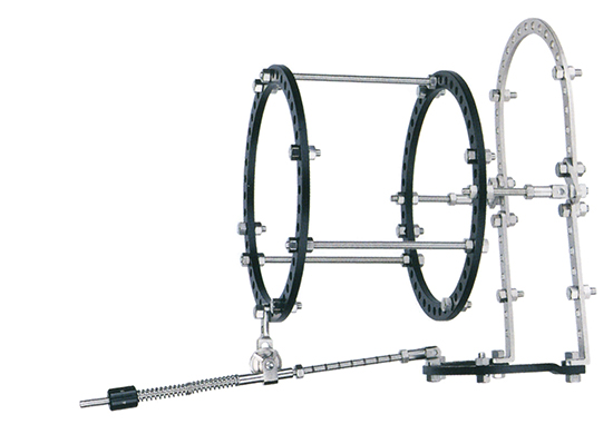
An external fixator plays a critical role in modern orthopedic trauma management, particularly when infection control becomes a primary clinical concern. This specialized device provides mechanical stability to fractured bones while simultaneously offering unique advantages for managing contaminated wounds and preventing surgical site infections. Understanding how an external fixator functions in infection control protocols is essential for surgical teams, hospital administrators, and medical device professionals involved in trauma care delivery.
The clinical value of an external fixator extends beyond simple fracture stabilization. When dealing with open fractures, contaminated wounds, or high-risk infection scenarios, an external fixator allows surgeons to keep the injury site accessible for repeated inspection, cleaning, and therapeutic intervention. This accessibility significantly reduces the time between injury and definitive treatment, a factor directly correlated with infection prevention outcomes in trauma literature.
How External Fixator Architecture Supports Infection Control
Minimizing Soft Tissue Trauma
An external fixator maintains bone alignment through percutaneous pins or wires that bypass extensive soft tissue disruption. Unlike internal fixation methods that require large incisions and extensive muscle stripping, an external fixator preserves vascular supply and tissue integrity. This preservation of soft tissue viability is a cornerstone of infection prevention, as compromised tissue perfusion creates an environment where bacteria proliferate unchecked. By reducing surgical trauma, an external fixator enables faster tissue healing and stronger innate immune response at the injury site.
Enabling Wound Access and Monitoring
One of the most valuable aspects of an external fixator in infection control is its design that leaves the wound site openly accessible. Surgeons can perform serial debridement, remove devitalized tissue, and inspect the wound without removing or repositioning the fracture fixation. An external fixator allows frequent dressing changes and direct observation of infection signs without disrupting bone stability. This continuous access becomes particularly valuable in polytrauma cases where multiple injuries require coordinated management and where infection monitoring must remain constant throughout recovery.
Clinical Protocols Using External Fixator for High-Risk Scenarios
Open Fracture Management Protocols
Open fractures carry inherently high infection risk due to direct bacterial contamination at the injury moment. An external fixator is the standard choice for temporary stabilization in open fracture protocols, allowing the surgical team to focus immediately on aggressive wound debridement and contamination control. Once contamination has been addressed and infection risk has declined over several days, surgeons may transition from temporary external fixator to definitive fixation. This staged approach, built around the unique advantages of external fixator accessibility, significantly improves infection outcomes compared to primary internal fixation in contaminated scenarios.
Soft Tissue Coverage and Reconstruction
When severe soft tissue injury accompanies fractures, an external fixator provides the stability necessary for plastic surgery intervention without compromising fracture alignment. The external fixator maintains skeletal stability while wound coverage specialists work on restoring soft tissue protection through flaps or grafts. An external fixator remains in place during these extended reconstructive procedures, ensuring that bone healing progresses without interruption even while infection prevention measures and tissue viability assessments continue. This coordination between orthopedic and plastic teams depends critically on the external fixator's ability to provide stable fixation without occupying the wound space.
Infection Control Advantages Over Internal Fixation Methods
Reduced Hardware-Related Biofilm Formation
Internal fixation implants, particularly plates and screws placed directly at the fracture site, can serve as biofilm substrates for bacterial colonization. An external fixator, by contrast, positions fixation elements outside the primary wound, significantly reducing the direct contact between implanted hardware and contaminated tissue. While pin sites do present their own infection risk, the percutaneous nature of an external fixator design allows for daily pin site care and visual monitoring that is simply impossible with buried hardware. This design difference makes an external fixator the preferred choice when infection prevention is the dominant clinical priority.
Simplified Revision and Replacement Pathways
If infection develops despite preventive efforts, an external fixator can be modified, adjusted, or replaced far more easily than internal fixation devices. Surgeons can exchange external fixator components, adjust frame geometry, or convert to an alternate external fixator configuration while maintaining bone alignment and wound accessibility. An external fixator's non-invasive nature means that modifications occur through percutaneous technique without creating new surgical wounds or deepening existing contamination. This flexibility is invaluable in complex trauma scenarios where infection management may require repeated interventions across extended treatment timelines.
FAQ
What types of fractures benefit most from external fixator use in infection control?
Open fractures, particularly Gustilo grade II and III injuries with significant soft tissue damage and contamination, are the primary candidates for external fixator stabilization in infection control protocols. Additionally, fractures in patients with severe comorbidities, immunosuppression, or vascular compromise benefit from the accessibility and reduced soft tissue trauma that an external fixator provides. Pelvic fractures with abdominal or perineal wounds frequently require external fixator stabilization to enable coordinated infection prevention across multiple wound areas. Each scenario prioritizes the external fixator's unique ability to maintain skeletal stability while keeping the injury site accessible for repeated intervention.
How long does an external fixator typically remain in place during infection management?
The timeline for external fixator use depends on contamination severity, soft tissue healing progression, and overall infection status. In aggressive open fracture protocols, an external fixator may serve as temporary fixation for seven to fourteen days while primary contamination control and serial debridement proceed. An external fixator might remain in place for several weeks in complex cases requiring extended wound surveillance or when soft tissue reconstruction demands prolonged stability. Surgeons reassess the infection status and tissue viability regularly, then transition from external fixator to definitive fixation once the clinical team has confidence that the infection risk has declined to acceptable levels for internal implantation.
What complications can occur at external fixator pin sites despite infection prevention efforts?
Pin site infections represent the most common complication associated with external fixator use, ranging from minor superficial cellulitis to deep pin tract osteomyelitis. Proper pin site care protocols, including daily cleansing, assessment, and antimicrobial management, significantly reduce but do not eliminate this risk. An external fixator pin site infection may require antibiotic therapy, pin removal and relocation, or in severe cases, conversion to alternate fixation methods. However, even when pin site infections occur, the overall infection profile of an external fixator remains favorable compared to internal fixation complications, and the external fixator's accessibility allows early detection and rapid intervention before deep bone involvement occurs.