
 EN
EN
In the evolving landscape of spinal surgery, precision is not merely a technical preference — it is a clinical imperative. The pedicle screw has become the cornerstone of modern spinal fixation systems, offering surgeons a reliable anchor point for stabilizing vertebrae across a wide range of pathologies. As minimally invasive spine surgery (MISS) continues to gain momentum for its patient-centered benefits, the demand for accurate pedicle screw placement has never been more significant. Any deviation from the intended trajectory can have immediate and long-term consequences for both surgical outcomes and patient safety.
Minimally invasive techniques fundamentally change how surgeons interact with spinal anatomy. Without the benefit of wide-field open exposure, every decision regarding pedicle screw trajectory must be informed by exceptional planning, advanced imaging, and purpose-built instrumentation. This article explores why accuracy in pedicle screw placement is so critical in minimally invasive procedures, examining the anatomical, biomechanical, clinical, and technological dimensions that together define the standard of care in contemporary spine surgery.
The Anatomical Complexity Behind Pedicle Screw Placement
Narrow Corridors and High-Stakes Margins
The pedicle is a narrow, bony bridge connecting the vertebral body to the posterior elements. Its dimensions vary significantly across spinal levels and individual patients, often measuring only a few millimeters in width at the thoracic spine. Placing a pedicle screw through this corridor without breaching the cortical walls requires a precise entry point, correct angulation, and controlled insertion depth. In open surgery, direct visualization helps compensate for some of these challenges, but in minimally invasive approaches, the tolerance for error is dramatically reduced.
Surrounding the pedicle are structures that leave no margin for imprecision. Medially lies the spinal cord or thecal sac. Laterally, vascular structures and nerve roots are at risk. Inferiorly and superiorly, disc spaces and adjacent neural elements create a confined zone of safe passage. A malpositioned pedicle screw — even by just a millimeter or two — can cause nerve injury, vascular damage, or dural tears that translate directly into patient morbidity. The stakes are highest in the thoracic region, where spinal cord proximity makes every degree of angulation consequential.
Patient-Specific Variability in Pedicle Anatomy
No two spines are the same, and this anatomical variability adds another layer of complexity to pedicle screw placement. Factors such as degenerative changes, scoliosis, osteoporosis, and previous surgical history can alter pedicle morphology significantly. Osteoporotic bone reduces the purchase strength of a pedicle screw, making initial accuracy even more critical because cortical breach in such cases may not be immediately apparent and can lead to delayed hardware failure. In deformed spines, standard anatomical landmarks may be unreliable, demanding image-guided approaches to ensure safe and effective screw positioning.
Preoperative CT imaging and surgical planning software now allow surgeons to study individual pedicle dimensions before making a single incision. This level of preparation is increasingly considered the standard of care, particularly in MIS contexts where intraoperative adjustments are more challenging. Understanding the anatomy of each patient's spine — rather than relying on population averages — is what separates acceptable outcomes from excellent ones when a pedicle screw is involved.
Biomechanical Implications of Accurate Pedicle Screw Positioning
Load Distribution and Construct Stability
The mechanical purpose of a pedicle screw is to transmit forces between the vertebra and the connecting rod or plate, enabling stabilization while fusion occurs. For this system to function as designed, each screw must engage the pedicle and vertebral body with adequate bone purchase. An accurately placed pedicle screw achieves bicortical or intracortical engagement that maximizes pullout strength. Conversely, a screw that breaches the medial wall or misses the pedicle entirely relies on weaker cancellous bone, compromising the entire construct.
In multi-level constructs, the cumulative effect of even small inaccuracies becomes pronounced. If several screws are slightly malpositioned, the resulting rod placement may introduce unintended forces into the spine, accelerating adjacent segment degeneration or creating asymmetric load distribution. Surgeons performing minimally invasive multilevel fusions must therefore treat each pedicle screw placement as part of an integrated mechanical system, where positional errors compound across levels rather than being isolated events.
Hardware Failure Prevention and Long-Term Outcomes
Accurate pedicle screw placement is directly linked to hardware longevity. Screws that are well-seated within the pedicle and vertebral body are less susceptible to the cyclical loading forces that can cause loosening, toggling, or fatigue fracture over time. In contrast, a malpositioned pedicle screw experiences abnormal stress concentrations at the bone-implant interface, often leading to premature failure. This can result in loss of fixation, need for revision surgery, and compromised fusion outcomes — all outcomes that are particularly undesirable given the patient populations typically treated with MIS techniques.
Patients who undergo minimally invasive spine surgery often do so specifically to achieve faster recovery and lower complication rates. Hardware failure undermines this expectation. Ensuring that each pedicle screw is placed with biomechanical precision is therefore not just a surgical quality metric — it is central to delivering on the clinical promise of minimally invasive approaches and maintaining patient confidence in the procedure and the care team.
How Minimally Invasive Approaches Amplify the Importance of Accuracy
Reduced Visual Field and the Challenge of Tactile Feedback
Open spine surgery allows surgeons to directly visualize the operative field, providing immediate feedback on tissue anatomy, screw trajectory, and construct alignment. Minimally invasive approaches sacrifice this panoramic view in exchange for smaller incisions, reduced muscle disruption, and faster patient recovery. The tradeoff means that surgeons must rely more heavily on fluoroscopy, navigation systems, and tactile sensing to ensure each pedicle screw is placed correctly. The challenge intensifies because small angular errors that would be immediately visible in open surgery can go undetected until imaging review in MIS procedures.
The tactile feedback available through MIS cannulas and dilators is also less informative than direct palpation. Surgeons must interpret subtle resistance changes during pedicle screw insertion as indicators of bone quality and wall integrity — a skill that requires significant experience and well-calibrated instrumentation. Purpose-designed MIS pedicle screw systems incorporate features such as cannulated construction, graduated depth markings, and sound-dampening handles that help surgeons interpret feedback more reliably even through the limited access portal.
Radiation Exposure and the Push Toward Navigation
Fluoroscopic guidance has long been the workhorse of MIS pedicle screw placement, offering real-time imaging feedback to confirm trajectory before and during screw insertion. However, reliance on fluoroscopy carries cumulative radiation exposure risks for both patients and surgical staff. As minimally invasive spine procedures increase in frequency, particularly for younger patient populations, the radiation burden associated with repeated fluoroscopic checks for each pedicle screw becomes a meaningful concern that the surgical community must address.
This has accelerated adoption of intraoperative 3D navigation and robotic assistance platforms, which can dramatically reduce fluoroscopy use while improving pedicle screw accuracy. Navigation-guided systems register the patient's preoperative or intraoperative CT data and provide the surgeon with real-time three-dimensional positional feedback for each instrument. Studies have consistently shown that navigation-assisted pedicle screw placement achieves higher accuracy rates compared to fluoroscopy-only techniques, with clinically significant reductions in breach rates and revision procedures. The integration of pedicle screw systems with advanced navigation platforms represents a significant step forward in making MIS procedures safer and more reproducible.
Clinical Consequences of Inaccurate Pedicle Screw Placement
Neurological Injury and Vascular Complications
The most serious consequence of a malpositioned pedicle screw is direct injury to neural or vascular structures. Medial breach of the pedicle wall can result in dural laceration, epidural hematoma, or direct nerve root or spinal cord compression. Depending on the spinal level and severity of breach, neurological consequences can range from transient radiculopathy to permanent motor deficits. These outcomes are among the most feared complications in spine surgery, and their potential for occurrence underscores why accuracy in pedicle screw placement must be treated as a non-negotiable standard.
Lateral breaches carry the risk of injuring segmental vessels, retroperitoneal structures, or nerve roots exiting the foramen. Anterior breaches, while less common, can cause devastating vascular injuries if a pedicle screw violates the anterior vertebral cortex and contacts the aorta or vena cava. The anatomical proximity of these structures demands that every pedicle screw be placed with awareness of all six cortical walls of the pedicle, not just the medial boundary that receives the most clinical attention.
Revision Surgery, Patient Recovery, and Healthcare Costs
Beyond neurological injury, inaccurate pedicle screw placement contributes significantly to revision surgery rates. When a malpositioned screw causes persistent pain, neurological symptoms, or construct instability, reoperation is often necessary. Revision spine surgery is inherently more complex, riskier, and more resource-intensive than the index procedure. For minimally invasive spine procedures specifically, where one of the primary goals is to minimize recovery time and complication burden, the need for revision surgery represents a fundamental failure to achieve the intended clinical outcome.
From a healthcare economics perspective, the cost implications of revision surgery are substantial. Longer hospital stays, additional implant costs, extended rehabilitation, and lost productivity all accumulate when a pedicle screw complication necessitates a return to the operating room. Investing in higher-accuracy instruments, navigation technology, and proper surgeon training for pedicle screw placement in MIS procedures is therefore not only a patient safety priority but also a rational economic strategy for healthcare systems aiming to optimize surgical value.
Instrumentation Design and Technological Innovation for MIS Pedicle Screw Accuracy
Cannulated Design and Guidewire-Based Systems
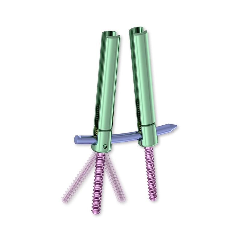
MIS-specific pedicle screw systems have been engineered to address the unique demands of limited-access surgery. Cannulated screw designs allow placement over a Kirschner wire or guidewire that has been precisely positioned under fluoroscopic or navigation guidance. This technique separates the trajectory-setting step from the screw insertion step, allowing the surgeon to confirm correct positioning before committing to hardware placement. The ability to verify and adjust the guidewire before inserting the pedicle screw is one of the most practically impactful safety features in modern MIS spine instrumentation.
Extender tubes and reduction towers integrated into pedicle screw systems for MIS applications also allow percutaneous rod passage through tissue corridors, maintaining screw head alignment during construct assembly. These components must be designed with dimensional accuracy to ensure that the mechanical relationships between individual pedicle screw heads remain consistent as the surgeon connects them with a rod — a task that requires precision engineering given that direct visualization of the connection point is limited in MIS procedures.
Material Science, Thread Design, and Bone Engagement
The clinical performance of a pedicle screw is not solely determined by where it is placed — it is also a function of how well it engages bone. Thread geometry, pitch, and outer diameter all influence pullout strength and resistance to toggling. For patients with reduced bone mineral density, specialized pedicle screw designs with expanded outer diameters, fenestrated shafts for cement augmentation, or dual-lead thread patterns can substantially improve fixation quality even when ideal bone stock is not available. These design innovations extend the clinical applicability of MIS fixation to patient populations that would previously have been considered poor candidates.
Titanium alloys remain the dominant material for pedicle screw systems due to their biocompatibility, strength-to-weight ratio, and compatibility with postoperative MRI imaging. Some newer systems incorporate cobalt-chromium alloys or PEEK components to modify construct stiffness profiles, though these materials each carry specific indications and tradeoffs. The material and design choices made by surgeons and hospitals when selecting a pedicle screw system have direct implications for clinical accuracy and patient outcomes, reinforcing the importance of evidence-based procurement decisions in the MIS surgical setting.
FAQ
What happens if a pedicle screw is placed inaccurately during MIS surgery?
An inaccurately placed pedicle screw can breach the pedicle wall and injure adjacent neural, vascular, or disc structures. The clinical consequences range from nerve root irritation and radiculopathy to more serious outcomes such as spinal cord injury or vascular damage. Beyond immediate injury, malpositioned hardware may compromise construct stability, accelerate hardware failure, and necessitate revision surgery. This is why accuracy is treated as the highest-priority parameter in pedicle screw placement during minimally invasive spine procedures.
How does navigation technology improve pedicle screw accuracy in MIS procedures?
Intraoperative navigation systems use CT data and real-time tracking to provide surgeons with three-dimensional positional feedback during pedicle screw insertion. This guidance reduces dependence on fluoroscopy and allows the surgeon to verify the screw trajectory against the patient's specific anatomy in real time. Navigation-assisted pedicle screw placement has demonstrated significantly lower pedicle breach rates compared to conventional fluoroscopic techniques, translating directly into fewer neurological complications and improved construct integrity.
Why is pedicle screw accuracy more challenging in minimally invasive surgery than in open surgery?
In open surgery, direct visualization of the surgical field provides continuous anatomical feedback that helps guide pedicle screw placement. Minimally invasive approaches use small incisions and tubular retractors that restrict the field of view, forcing surgeons to rely on imaging guidance and tactile feedback rather than direct visualization. This restricted access means that errors in pedicle screw trajectory are harder to detect intraoperatively and may only become apparent upon fluoroscopic review. Specialized MIS instrumentation and navigation technology are therefore essential tools for maintaining accuracy under these constrained conditions.
What role does preoperative planning play in ensuring accurate pedicle screw placement?
Preoperative CT-based planning allows surgeons to assess each patient's unique pedicle dimensions, morphology, and bone quality before surgery begins. This information guides the selection of pedicle screw size and length, determines safe entry points and angulation, and identifies anatomical variants that could increase procedural risk. In complex cases involving deformity or osteoporosis, detailed preoperative planning is especially critical to ensuring that each pedicle screw can be placed accurately and that the construct will perform as intended over the long term. Planning software that interfaces with intraoperative navigation further strengthens the continuity between surgical planning and execution.
Table of Contents
- The Anatomical Complexity Behind Pedicle Screw Placement
- Biomechanical Implications of Accurate Pedicle Screw Positioning
- How Minimally Invasive Approaches Amplify the Importance of Accuracy
- Clinical Consequences of Inaccurate Pedicle Screw Placement
- Instrumentation Design and Technological Innovation for MIS Pedicle Screw Accuracy
-
FAQ
- What happens if a pedicle screw is placed inaccurately during MIS surgery?
- How does navigation technology improve pedicle screw accuracy in MIS procedures?
- Why is pedicle screw accuracy more challenging in minimally invasive surgery than in open surgery?
- What role does preoperative planning play in ensuring accurate pedicle screw placement?