
When a patient sustains a serious bone fracture or joint injury, the goal of trauma surgery is to restore stability, alignment, and function as efficiently and safely as possible. Central to achieving that goal are orthopedic implants — precision-engineered devices designed to support, fixate, or replace damaged skeletal structures. Understanding the main types of orthopedic implants used in trauma surgery is essential not only for surgeons and medical professionals, but also for procurement teams, hospital administrators, and biomedical engineers who are responsible for sourcing and stocking the right instruments and hardware.
The landscape of orthopedic implants has evolved dramatically over the past several decades. Advances in metallurgy, biomechanics, and surgical technique have produced a wide and specialized range of fixation devices and reconstruction solutions. Each category of implant is engineered with a specific type of fracture pattern, anatomical location, or biomechanical challenge in mind. This article breaks down the primary implant families used in trauma surgery, explains their clinical logic, and highlights the technical distinctions that make each type uniquely suited to particular trauma scenarios.
Bone Plates and Screws
The Foundation of Internal Fixation
Bone plates and screws represent one of the most widely used categories of orthopedic implants in trauma surgery. These devices work by providing rigid or semi-rigid fixation across a fracture site, holding bone fragments in their correct anatomical position while the healing process occurs. Plates are typically affixed to the bone surface using cortical or cancellous screws, and they come in a variety of shapes and sizes to accommodate different bones and fracture configurations.
The mechanical principle behind plate-and-screw systems is compression or bridging, depending on the fracture type. In a simple transverse fracture, a compression plate can actively press the two fragments together, creating ideal conditions for direct bone healing. In comminuted fractures where multiple fragments are present, a bridging plate spans the zone of injury without disturbing the fragments, allowing indirect healing through callus formation.
From a materials standpoint, most modern plates are fabricated from titanium alloy or stainless steel, both of which offer high strength-to-weight ratios and favorable biocompatibility profiles. The choice of material often depends on the specific surgical indication, patient profile, and whether the implant is intended for permanent retention or planned removal after healing.
Locking Plate Technology in Trauma
A significant advancement within the plate-and-screw family is the development of locking plates, which feature threaded screw holes that allow screws to lock into the plate at a fixed angle. This locking mechanism creates a fixed-angle construct, effectively turning the plate and screws into a single load-sharing unit. Locking technology is particularly valuable in osteoporotic bone, where conventional screw purchase may be insufficient to maintain fracture reduction.
The orthopedic implants available in small fragment locking sets are a prime example of how this technology has been refined for distal radius fractures, hand and wrist injuries, and pediatric cases where bone diameter is reduced. These sets typically include a curated collection of plates, locking screws, cortical screws, and the associated instrumentation needed to perform precise fixation.
Locking plates also allow surgeons to maintain a degree of biological fixation — meaning the plate does not need to compress tightly against the periosteum, preserving blood supply and reducing the risk of thermal necrosis and delayed union. This biological advantage has made locking plate systems a preferred choice in many modern trauma centers around the world.
Intramedullary Nails
Axial Load-Sharing in Long Bone Fractures
Intramedullary (IM) nails are orthopedic implants inserted directly into the medullary canal of long bones such as the femur, tibia, and humerus. Unlike plates, which sit on the bone surface, IM nails occupy the central axis of the bone, making them inherently more aligned with the physiological load-bearing axis. This biomechanical positioning allows the nail to share axial loads with the bone rather than bearing them entirely, which promotes more natural healing mechanics.
IM nails are inserted using a minimally invasive technique, often through a small entry portal at one end of the bone. Interlocking screws are then placed through the nail at both the proximal and distal ends to control rotation and prevent shortening. This locked nail construct is the standard of care for many diaphyseal fractures because it combines reliable fixation with minimal soft tissue disruption.
The clinical advantages of IM nailing are particularly pronounced in femoral shaft fractures, where the load demands are highest and where early mobilization is critical to preventing complications such as deep vein thrombosis and pneumonia. The ability to begin weight-bearing soon after surgery is a major benefit that IM nails offer over plate-based alternatives in many long bone scenarios.
Nail Design Variations and Clinical Selection
IM nails are not a single uniform device. They come in a variety of configurations designed for specific bones and fracture locations. Antegrade nails are inserted from the proximal end of the bone, while retrograde nails are inserted from the distal end. This distinction matters significantly for fracture patterns near joints, where one approach may preserve articular integrity better than the other.
Cephalomedullary nails incorporate a proximal blade or screw that extends into the femoral head, making them the implant of choice for intertrochanteric and subtrochanteric hip fractures. These nails combine the load-sharing properties of an IM device with the rotational control needed to stabilize periarticular fractures in the proximal femur.
Material selection for IM nails follows similar considerations as plates — titanium is preferred in cases where MRI compatibility is a concern or where implant retention is expected to be long-term. Nail diameter and length must be carefully templated preoperatively using imaging to ensure proper fit within the medullary canal without causing cortical stress concentrations at the nail tips.
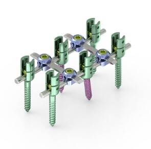
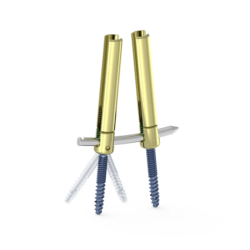
External Fixators
Temporary and Definitive Fixation Outside the Body
External fixators represent a category of orthopedic implants that provide fracture stabilization from outside the skin. Unlike plates and nails, which are fully implanted, external fixators use percutaneous pins or wires that pass through the skin and bone and connect to an external frame. This design makes them ideal in situations where internal fixation would be unsafe or impractical.
Common indications for external fixation in trauma include open fractures with significant soft tissue contamination, highly comminuted fractures requiring spanning of a joint, damage control orthopedics in polytrauma patients, and cases where definitive surgery must be delayed due to systemic instability. In these scenarios, an external fixator can rapidly restore length, alignment, and rotation without exposing the wound zone to hardware.
Modern external fixators use modular carbon fiber or aluminum frames that allow the surgeon to adjust alignment postoperatively, which can be a significant advantage in managing complex periarticular injuries. Some systems are designed to transition into definitive fixation after soft tissue recovery, at which point the frame is replaced with internal hardware.
Circular and Ring Fixators for Complex Cases
Circular external fixators, such as the Ilizarov frame and its derivatives, use a series of rings connected by threaded rods and tensioned wires to achieve highly stable fixation with minimal bone contact. These systems are particularly valuable in the management of nonunions, malunions, bone transport after segmental defects, and complex periarticular injuries where fine, multi-planar correction is required.
The biomechanical principles of circular fixation differ significantly from conventional uniplanar external fixation. The fine wires, when tensioned appropriately, create a stable fixation construct that can tolerate axial micromotion, which has been shown to stimulate callus formation and promote fracture healing. This controlled micromotion is in contrast to the rigidity of internal fixation methods, and it represents a deliberately different healing strategy.
External fixators in all their forms are important orthopedic implants within the trauma surgeon's toolkit, bridging the gap between emergency stabilization and definitive reconstruction. Their versatility, combined with the ability to perform outside conventional implant pathways, gives them an enduring role even as internal fixation technology continues to advance.
Cannulated Screws and K-Wires
Small but Critical Fixation Devices
Cannulated screws and Kirschner wires (K-wires) are among the smallest yet most frequently employed orthopedic implants in trauma surgery. Cannulated screws are hollow and designed to be inserted over a guidewire, allowing precise targeting under fluoroscopic guidance. They are particularly effective for percutaneous fixation of small fractures, epiphyseal injuries in children, and fractures in anatomically confined regions such as the scaphoid, femoral neck, and calcaneus.
The minimally invasive nature of cannulated screw fixation makes it well-suited to fractures in well-vascularized cancellous bone, where the compression applied by the screw is sufficient to maintain reduction without additional implants. In femoral neck fractures, three cannulated screws arranged in a triangular pattern provide rotational stability while allowing the sliding compression needed for impaction healing.
K-wires are smooth, thin, pointed metal wires that can be used for temporary fixation during surgery or as a definitive fixation strategy for small bone fragments. They are inexpensive, easy to insert and remove, and available in a range of diameters. In pediatric trauma, K-wires are a primary fixation tool due to their compatibility with growing bone and their removability without significant secondary trauma.
Tension Band Wiring and Augmented Screw Constructs
In certain anatomical locations, K-wires are combined with cerclage wire in a technique known as tension band wiring. This construct converts the distracting forces acting on an avulsion fragment — such as at the olecranon or patella — into compressive forces at the fracture site. Tension band constructs are among the most biomechanically elegant solutions in trauma surgery, converting muscle pull into healing compression.
Cannulated screws can also be used in combination with other orthopedic implants to augment fixation in complex reconstructions. For example, a cannulated screw may be used alongside a buttress plate to prevent rotation of a proximal tibial fracture fragment, or in combination with bone graft to support fixation in osteoporotic bone with poor screw purchase.
The variety of sizes, thread designs, and cannulation diameters available today reflects the high level of customization now possible in trauma fixation. Matching the right screw geometry to the specific bone density, fracture morphology, and healing expectations of each patient is a nuanced skill that distinguishes expert trauma surgeons and well-equipped operating theaters.
Joint Replacement Implants in Acute Trauma
When Reconstruction Is Not Enough
In some trauma scenarios, the degree of articular damage or bone loss is too severe for conventional fixation strategies. In these cases, partial or total joint replacement becomes the appropriate intervention. Arthroplasty implants in the context of trauma — as opposed to elective joint replacement for arthritis — must meet a slightly different set of demands, including compatibility with emergency surgical settings, availability in a range of sizes, and durability in patients who may be physiologically stressed.
Hemiarthroplasty of the femoral head is a classic example of an arthroplasty application in acute trauma. In displaced femoral neck fractures in elderly patients, the risk of avascular necrosis following internal fixation makes prosthetic replacement a more reliable option. A femoral head prosthesis restores limb length and joint function while bypassing the biological unpredictability of healing in compromised bone.
These orthopedic implants are designed to integrate with existing anatomical structures — typically the acetabulum in hip cases — without requiring full reconstruction of both joint surfaces. The ability to mobilize patients quickly after hemiarthroplasty is a major clinical benefit, especially in elderly populations where prolonged immobility carries significant risks of cardiopulmonary and thromboembolic complications.
Total Elbow and Shoulder Replacement in Trauma
Highly comminuted fractures of the distal humerus or proximal humerus in elderly patients represent scenarios where total elbow or shoulder arthroplasty may be the most practical solution. The fracture pattern in these cases often makes anatomical reconstruction virtually impossible, and the expected bone quality may not support the multiple screws and plates needed for rigid fixation.
Reverse total shoulder arthroplasty has gained significant traction in trauma settings for proximal humeral fractures in elderly patients, offering reliable functional outcomes even when the rotator cuff is compromised or the tuberosities cannot be reliably fixed. This design inverts the normal ball-and-socket geometry to shift the center of rotation medially, compensating for rotator cuff deficiency and relying instead on the deltoid muscle for elevation.
The use of arthroplasty-based orthopedic implants in trauma requires careful patient selection, preoperative planning, and access to appropriately sized modular systems. Surgical teams must be equipped with a range of stem lengths, head offsets, and glenosphere or acetabular cup sizes to accommodate the anatomical variation encountered in emergency cases.
FAQ
What is the most commonly used orthopedic implant in trauma surgery?
Bone plates and screws, including locking plate systems, are among the most frequently used orthopedic implants in trauma surgery due to their versatility across a wide range of fracture types and anatomical locations. Intramedullary nails are also widely used, particularly for diaphyseal fractures of the femur and tibia. The 'most common' implant depends heavily on the anatomical region and fracture pattern being treated.
How are locking plates different from standard bone plates?
Locking plates differ from standard bone plates in that their screw holes have internal threads that engage corresponding threads on the screw head, creating a fixed-angle locked construct. Standard plates rely on friction between the plate and the bone surface to maintain stability, while locking plates create a rigid mechanical unit between the plate and screws. This makes locking plates significantly more effective in osteoporotic bone and in periarticular fracture regions where screw purchase is limited.
When are external fixators preferred over internal fixation?
External fixators are preferred when internal fixation poses unacceptable risks, such as in open fractures with contaminated soft tissue environments, in polytrauma patients requiring damage control surgery, or in cases where significant swelling makes wound closure over internal hardware unsafe. They are also used when definitive surgical reconstruction must be delayed due to the patient's systemic condition or when complex deformity correction requiring gradual adjustment is needed.
Can orthopedic implants be removed after bone healing?
Many orthopedic implants can be removed after fracture healing, although the decision to remove them depends on several factors including patient symptoms, implant location, patient age, and whether the implant is causing soft tissue irritation or interfering with joint function. In children, implant removal is often planned as part of the treatment protocol to avoid interference with skeletal growth. In adults, asymptomatic implants are often left in place permanently, while prominently positioned hardware that causes discomfort may be removed electively after confirmed healing.