
Orthopedic trauma surgery has evolved significantly over the past decades, with various fixation methods available to surgeons treating complex fractures. Among these options, the interlocking nail stands out as a revolutionary approach that has transformed the management of long bone fractures. This advanced fixation technique combines the biomechanical advantages of intramedullary nailing with enhanced stability through proximal and distal locking mechanisms, making it an invaluable tool in modern orthopedic practice.
Understanding Interlocking Nail Technology
Biomechanical Principles of Interlocking Systems
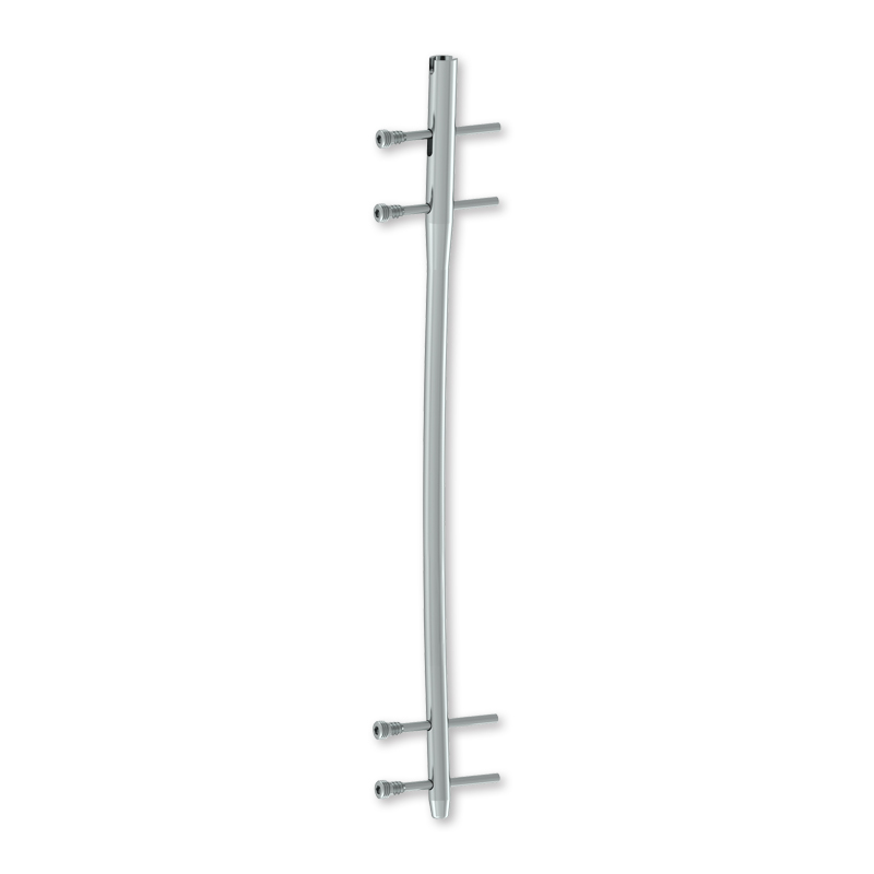
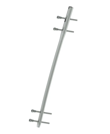
The fundamental design of an interlocking nail incorporates sophisticated engineering principles that address the complex mechanical demands of fractured long bones. Unlike traditional intramedullary rods, these devices feature multiple holes positioned strategically along the nail's length, allowing surgeons to insert locking screws that secure the implant to the bone. This design creates a rigid construct that resists rotational forces, prevents shortening, and maintains proper alignment throughout the healing process.
The material composition of modern interlocking nails typically involves titanium alloys or stainless steel, each offering distinct advantages in terms of biocompatibility, strength, and imaging compatibility. The nail's cannulated design facilitates insertion over a guide wire, reducing surgical trauma while ensuring precise placement within the medullary canal. Advanced surface treatments and coatings further enhance osseointegration and reduce the risk of infection.
Anatomical Considerations for Nail Selection
Proper selection of an interlocking nail requires thorough understanding of the patient's anatomy and the specific characteristics of the fracture pattern. Femoral applications represent the most common use case, where the nail's design must accommodate the natural curvature of the femur while providing adequate fixation strength. The diameter and length selection process involves careful measurement of the medullary canal and consideration of the patient's bone quality and activity level.
Tibial interlocking nails present unique challenges due to the bone's triangular cross-section and the presence of the fibula. Surgeons must consider the entry point, typically through the tibial plateau, and ensure that the nail's distal end doesn't interfere with ankle function. The locking screw configuration becomes particularly critical in preventing malrotation and maintaining proper alignment of the tibiofibular relationship.
Clinical Indications and Patient Selection
Fracture Patterns Best Suited for Interlocking Fixation
Unstable long bone fractures represent the primary indication for interlocking nail fixation, particularly those involving the femoral and tibial shafts. Comminuted fractures, where multiple bone fragments create inherent instability, benefit significantly from the rigid fixation provided by the interlocking mechanism. Segmental fractures, characterized by separate fracture lines creating a floating bone segment, require the longitudinal support and rotational control that only an interlocking nail can provide effectively.
Pathological fractures through metastatic lesions or osteoporotic bone present another compelling indication for interlocking nail use. The device's ability to span large defects while providing immediate stability makes it ideal for patients with compromised bone quality. Additionally, fractures in the subtrochanteric region of the femur, known for their challenging biomechanical environment, often require the enhanced fixation strength that interlocking nail systems provide to prevent implant failure.
Patient Factors Influencing Treatment Decisions
Age and activity level play crucial roles in determining the appropriateness of interlocking nail fixation. Younger, more active patients typically benefit from the immediate weight-bearing potential that these devices offer, allowing for faster return to normal activities. Elderly patients with osteoporotic bone may require specialized nail designs with enhanced locking mechanisms to compensate for reduced bone quality and prevent screw cutout.
Comorbidities such as diabetes, peripheral vascular disease, or immunocompromising conditions require careful consideration when selecting fixation methods. The minimally invasive nature of interlocking nail insertion often makes it preferable to extensive open reduction techniques in medically compromised patients. However, the surgeon must balance the benefits of reduced surgical exposure against the technical demands of achieving proper reduction and nail placement.
Surgical Technique and Procedural Excellence
Preoperative Planning and Imaging Requirements
Successful interlocking nail insertion begins with meticulous preoperative planning that includes comprehensive imaging studies and templating. High-quality anteroposterior and lateral radiographs provide essential information about fracture configuration, bone quality, and medullary canal dimensions. Advanced imaging modalities such as CT scans may be necessary for complex fracture patterns or when planning revision procedures.
Template overlay techniques help surgeons select appropriate nail diameter and length while identifying potential technical challenges. The presence of existing hardware, previous surgical interventions, or anatomical variants must be carefully evaluated during the planning phase. Surgeons should also consider the availability of specialized instrumentation and backup plans for potential complications during the procedure.
Intraoperative Techniques for Optimal Outcomes
The surgical approach for interlocking nail insertion typically involves a small incision over the entry point, minimizing soft tissue disruption compared to traditional open reduction techniques. Proper patient positioning on a fracture table or with manual traction becomes critical for achieving and maintaining reduction throughout the procedure. The use of fluoroscopic guidance ensures accurate nail placement and proper positioning of locking screws.
Reduction techniques may involve closed manipulation, percutaneous reduction aids, or limited open approaches depending on fracture complexity. The insertion of the interlocking nail requires careful attention to rotation, length, and the relationship between proximal and distal fragments. Locking screw placement demands precision to avoid neurovascular structures while ensuring adequate purchase in cortical bone for maximum fixation strength.
Comparative Analysis with Alternative Fixation Methods
Advantages Over Plate and Screw Constructs
When compared to traditional plate and screw fixation, interlocking nails offer several distinct biomechanical advantages that make them superior choices for specific fracture patterns. The load-sharing characteristics of intramedullary fixation distribute forces more physiologically compared to the load-bearing nature of plate constructs. This fundamental difference reduces the risk of implant failure and promotes more natural bone healing patterns.
The minimally invasive insertion technique preserves the fracture hematoma and reduces soft tissue stripping, factors that contribute to enhanced healing potential. Additionally, the smaller surgical exposure translates to reduced operative time, decreased blood loss, and lower infection rates. The cosmetic advantages of smaller incisions also contribute to improved patient satisfaction and reduced long-term morbidity.
Limitations and Contraindications
Despite their numerous advantages, interlocking nails have specific limitations that surgeons must recognize when making treatment decisions. Fractures extending into the articular surfaces typically require additional fixation methods or alternative approaches to address joint congruity. Very proximal or distal fractures may not have adequate bone stock for effective locking screw placement, limiting the applicability of this technique.
Technical challenges associated with interlocking nail insertion include the learning curve required for proficient use of targeting systems and fluoroscopic guidance. Malreduction or improper nail placement can result in complications such as malunion, nonunion, or hardware failure. Additionally, certain fracture patterns with significant comminution may require supplemental fixation techniques to achieve optimal stability.
Long-term Outcomes and Follow-up Considerations
Healing Patterns and Bone Remodeling
The healing response following interlocking nail fixation typically follows predictable patterns that surgeons can monitor through serial radiographic evaluation. Secondary bone healing, characterized by callus formation and gradual remodeling, represents the normal response to stable fixation with controlled motion at the fracture site. The interlocking nail's design allows for some degree of dynamization as healing progresses, promoting natural bone remodeling processes.
Factors influencing healing rates include patient age, smoking status, nutritional state, and compliance with weight-bearing restrictions. The majority of fractures treated with interlocking nails achieve union within three to six months, with radiographic evidence of bridging callus and clinical signs of healing. Delayed union or nonunion may require additional interventions such as dynamization, bone grafting, or exchange nailing procedures.
Implant Removal Considerations
The question of implant removal following successful fracture healing remains a topic of ongoing debate in orthopedic surgery. Many interlocking nails can remain in place permanently without causing significant complications, particularly in older patients or those with lower activity demands. However, younger patients may benefit from nail removal to restore normal bone mechanics and eliminate the risk of long-term implant-related complications.
Indications for nail removal include persistent pain, interference with activities, or patient preference after thorough discussion of risks and benefits. The removal procedure typically involves extraction of locking screws followed by nail removal, though technical challenges may arise due to bone ingrowth or implant integration. Surgeons should counsel patients about the potential for complications during removal procedures, including fracture risk and the need for activity modifications during the healing period.
FAQ
What makes an interlocking nail different from a regular intramedullary rod?
An interlocking nail differs from a standard intramedullary rod through its sophisticated locking mechanism that includes proximal and distal screw holes. While regular rods provide longitudinal support, interlocking nails add rotational stability and prevent shortening through cross-locking screws that secure the implant to the bone. This enhanced stability makes interlocking nails ideal for unstable fracture patterns that would be inadequately controlled with simple rodding techniques.
How long does recovery typically take with interlocking nail fixation?
Recovery time following interlocking nail insertion varies depending on fracture complexity, patient factors, and compliance with rehabilitation protocols. Most patients can begin partial weight-bearing within the first few weeks, progressing to full weight-bearing as healing progresses. Complete bone healing typically occurs within three to six months, with return to normal activities possible once radiographic union is confirmed and strength is restored through physical therapy.
Are there any long-term complications associated with interlocking nails?
Long-term complications following interlocking nail fixation are relatively uncommon but can include chronic pain, implant failure, or issues related to locking screws. Some patients may experience knee pain with femoral nails due to the entry point through the piriformis fossa. Hardware removal may be considered for persistent symptoms, though many patients function well with permanent implants. Regular follow-up allows for early detection and management of any developing complications.
Can interlocking nails be used in patients with osteoporosis?
Interlocking nails can be successfully used in osteoporotic patients, though special considerations apply regarding screw placement and bone quality. Modern nail designs include features such as angular stable locking and cement augmentation options to enhance fixation in compromised bone. The load-sharing nature of intramedullary fixation often makes it preferable to plate constructs in osteoporotic bone, as it reduces stress concentration and the risk of implant cutout through weakened cortices.