
Precision in orthopedic surgery demands meticulous attention to detail, particularly when performing cannulated screw placement procedures. Modern surgical techniques have evolved to incorporate advanced imaging technologies, specialized instrumentation, and refined methodologies that significantly enhance placement accuracy. Surgeons worldwide recognize that optimal patient outcomes depend heavily on the precise positioning of cannulated screws, which serve as critical fixation devices in fracture repair and reconstructive procedures. The integration of real-time imaging guidance, proper patient positioning, and standardized surgical protocols has revolutionized how medical professionals approach these complex interventions.
Imaging-Guided Navigation Systems
Fluoroscopic Real-Time Monitoring
Fluoroscopic guidance represents the gold standard for real-time visualization during cannulated screw insertion procedures. This imaging modality provides continuous monitoring capabilities that allow surgeons to observe guidewire trajectory and screw advancement throughout the entire procedure. Advanced fluoroscopic systems offer enhanced image quality with reduced radiation exposure, making them safer for both patients and surgical teams. The integration of multiple viewing angles ensures comprehensive visualization of anatomical structures and implant positioning.
Contemporary fluoroscopic equipment incorporates digital enhancement features that improve bone-to-soft tissue contrast ratios. These technological improvements enable surgeons to identify critical anatomical landmarks with greater precision, reducing the risk of neurovascular complications. Modern systems also provide measurement tools that facilitate accurate screw length selection and trajectory planning before actual insertion begins.
Three-Dimensional Imaging Integration
Three-dimensional imaging technologies have transformed preoperative planning and intraoperative guidance for cannulated screw procedures. CT-based navigation systems create detailed anatomical models that surgeons can reference during surgery, providing unprecedented accuracy in screw trajectory planning. These systems calculate optimal entry points and angulation based on patient-specific anatomy, significantly reducing placement errors.
Intraoperative CT scanning capabilities allow for immediate verification of screw positioning before wound closure. This real-time feedback mechanism enables surgeons to make necessary adjustments during the procedure, eliminating the need for revision surgeries due to malpositioned implants. The combination of preoperative planning and intraoperative verification creates a comprehensive quality control system that maximizes placement accuracy.
Surgical Technique Optimization
Guidewire Placement Strategies
Precise guidewire insertion forms the foundation of accurate cannulated screw placement, requiring careful consideration of anatomical landmarks and trajectory planning. Surgeons must identify optimal entry points that provide the most direct path to target locations while avoiding critical neurovascular structures. The use of multiple guidewires allows for triangulation techniques that enhance stability and distribute mechanical loads more effectively across fracture sites.
Parallel guidewire placement techniques have gained popularity due to their ability to provide uniform compression across fracture planes. This method requires specialized instrumentation and precise angle control to ensure that all guidewires maintain appropriate spacing and orientation. Surgeons often employ targeting devices that maintain consistent angles between multiple guidewires, reducing the complexity of parallel placement procedures.
Drilling and Measurement Protocols
Systematic measurement protocols ensure that cannulated screws achieve optimal purchase in target bone segments. Depth gauges specifically designed for cannulated systems provide accurate length measurements that account for guidewire diameter and screw thread engagement. These measurements must consider cortical thickness variations and cancellous bone density differences that affect screw holding power.
Controlled drilling techniques minimize thermal necrosis and preserve bone integrity around screw insertion sites. Intermittent drilling with frequent irrigation helps maintain acceptable temperatures while removing bone debris that could interfere with screw insertion. Modern drilling systems incorporate torque-limiting features that prevent excessive force application during bone preparation phases.
Patient Positioning and Anatomical Considerations
Optimal Patient Positioning Techniques
Strategic patient positioning directly influences the accessibility and accuracy of cannulated screw placement procedures. Proper positioning ensures that imaging equipment can provide clear visualization from multiple angles while maintaining sterile surgical fields. Surgeons must consider both primary surgical access requirements and secondary imaging needs when determining optimal patient positioning.
Specialized positioning devices and radiolucent table accessories facilitate consistent patient placement across multiple cases. These tools help maintain anatomical alignment while allowing unobstructed imaging access throughout the procedure. The use of standardized positioning protocols reduces setup time and improves overall surgical efficiency while maintaining safety standards.
Anatomical Landmark Recognition
Accurate identification of anatomical landmarks serves as the cornerstone of successful cannulated screw procedures. Surgeons must develop proficiency in recognizing key bony prominences, joint spaces, and soft tissue boundaries that guide screw placement decisions. Palpation techniques combined with imaging correlation help establish reliable reference points for surgical navigation.
Understanding regional anatomical variations becomes crucial when treating diverse patient populations with different bone morphologies. Age-related changes, pathological conditions, and previous surgical interventions can alter normal anatomical relationships, requiring adaptive surgical approaches. Comprehensive preoperative imaging review helps identify these variations and adjust surgical plans accordingly.
Quality Control and Verification Methods
Intraoperative Assessment Techniques
Comprehensive intraoperative assessment protocols ensure that cannulated screw placement meets established accuracy standards before surgical completion. Multiple imaging views from different angles provide complete evaluation of screw position relative to anatomical structures and fracture alignment. Surgeons employ standardized checklists that verify critical placement parameters including screw length, thread engagement, and trajectory alignment.
Real-time compression testing allows surgeons to evaluate fracture stability and screw purchase immediately after insertion. These assessments help identify inadequate fixation before wound closure, enabling corrective measures when necessary. Advanced monitoring systems can quantify compression forces and provide objective measurements of fixation quality.
Post-Placement Imaging Protocols
Systematic post-placement imaging verification confirms optimal screw positioning and identifies potential complications before they become clinically significant. High-resolution radiographic images from multiple projections document final implant positions for comparison with preoperative plans. These images serve as baseline references for subsequent follow-up evaluations and surgical outcome assessments.
Digital imaging systems enable immediate image processing and analysis that can detect subtle positioning errors not visible during intraoperative fluoroscopy. Automated measurement tools help quantify screw angles, lengths, and spacing with greater precision than traditional manual methods. These technological capabilities support evidence-based decision-making regarding the need for immediate revision procedures.
Instrumentation and Equipment Selection
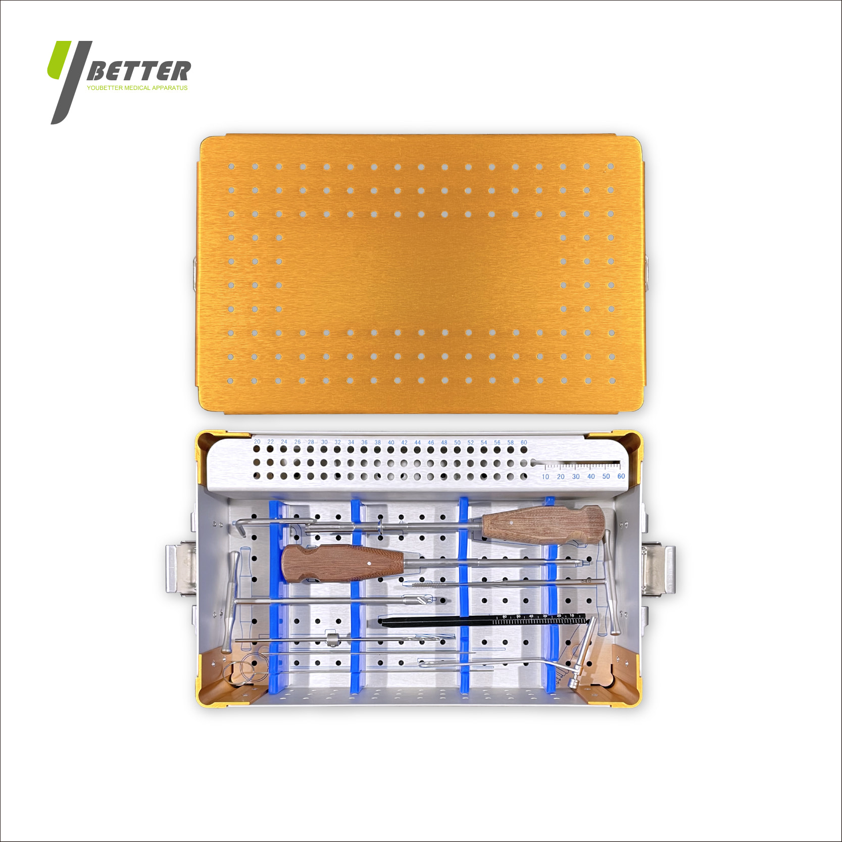
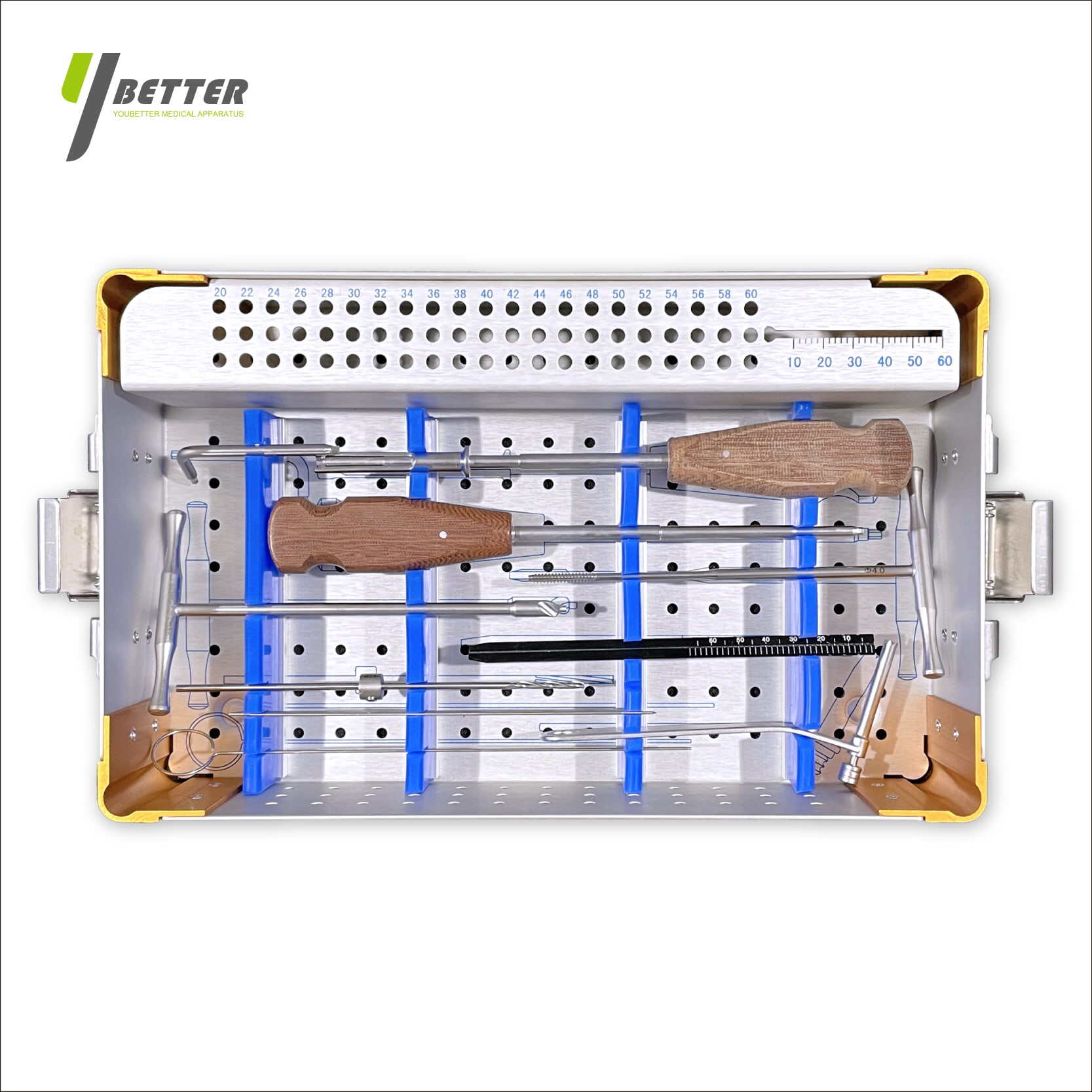
Specialized Cannulated Screw Systems
Modern cannulated screw systems incorporate design features that enhance placement accuracy and reduce procedural complexity. Self-drilling cannulated screws eliminate separate drilling steps while maintaining precise trajectory control through integrated guidewire channels. These systems often include depth-limiting features that prevent over-advancement and reduce the risk of cortical breach on the far side of target bones.
Variable pitch thread designs optimize screw purchase in different bone densities while providing controlled compression across fracture sites. Headless screw designs minimize soft tissue irritation while maintaining strong fixation capabilities. The selection of appropriate screw diameter and length depends on bone quality assessments and mechanical loading requirements specific to each anatomical location.
Targeting and Alignment Devices
Precision targeting devices help maintain consistent cannulated screw placement angles and reduce operator-dependent variability. These instruments incorporate adjustable guides that can be customized for different anatomical locations and surgical approaches. Some systems include laser alignment features that provide visual trajectory confirmation before guidewire insertion begins.
Computer-assisted targeting systems combine preoperative imaging data with intraoperative navigation to provide real-time guidance throughout the procedure. These systems calculate optimal screw trajectories based on patient-specific anatomy and fracture patterns, reducing reliance on surgeon experience alone. The integration of haptic feedback mechanisms provides tactile confirmation of proper instrument positioning and alignment.
FAQ
What imaging techniques provide the most accurate guidance for cannulated screw placement?
Fluoroscopic guidance combined with three-dimensional CT navigation systems offers the highest accuracy for cannulated screw placement procedures. Real-time fluoroscopy provides continuous visualization during insertion, while 3D imaging enables precise preoperative planning and intraoperative verification. The combination of multiple imaging modalities creates a comprehensive guidance system that significantly reduces placement errors and improves patient outcomes.
How do surgeons ensure proper guidewire placement before screw insertion?
Surgeons use multiple fluoroscopic views and anatomical landmark identification to confirm optimal guidewire positioning before proceeding with screw insertion. Parallel guidewire techniques often employ specialized targeting devices that maintain consistent spacing and angulation. Depth measurements and trajectory verification through multiple imaging planes ensure that guidewires follow planned paths and avoid critical anatomical structures.
What factors influence the selection of cannulated screw length and diameter?
Screw length selection depends on cortical thickness measurements, bone density assessments, and the need for adequate thread engagement in target bone segments. Diameter selection considers the mechanical loading requirements of the specific anatomical location and the size of the medullary canal or fracture fragments. Preoperative imaging analysis and intraoperative measurements help determine optimal screw specifications for each individual case.
How can surgeons minimize complications during cannulated screw procedures?
Complication prevention relies on thorough preoperative planning, precise surgical technique, and comprehensive intraoperative monitoring. Surgeons should utilize multiple imaging views, follow standardized placement protocols, and verify screw position before wound closure. Proper patient positioning, appropriate instrumentation selection, and adherence to sterile technique principles contribute to successful outcomes and reduced complication rates.
Table of Contents
- Imaging-Guided Navigation Systems
- Surgical Technique Optimization
- Patient Positioning and Anatomical Considerations
- Quality Control and Verification Methods
- Instrumentation and Equipment Selection
-
FAQ
- What imaging techniques provide the most accurate guidance for cannulated screw placement?
- How do surgeons ensure proper guidewire placement before screw insertion?
- What factors influence the selection of cannulated screw length and diameter?
- How can surgeons minimize complications during cannulated screw procedures?