
 EN
EN
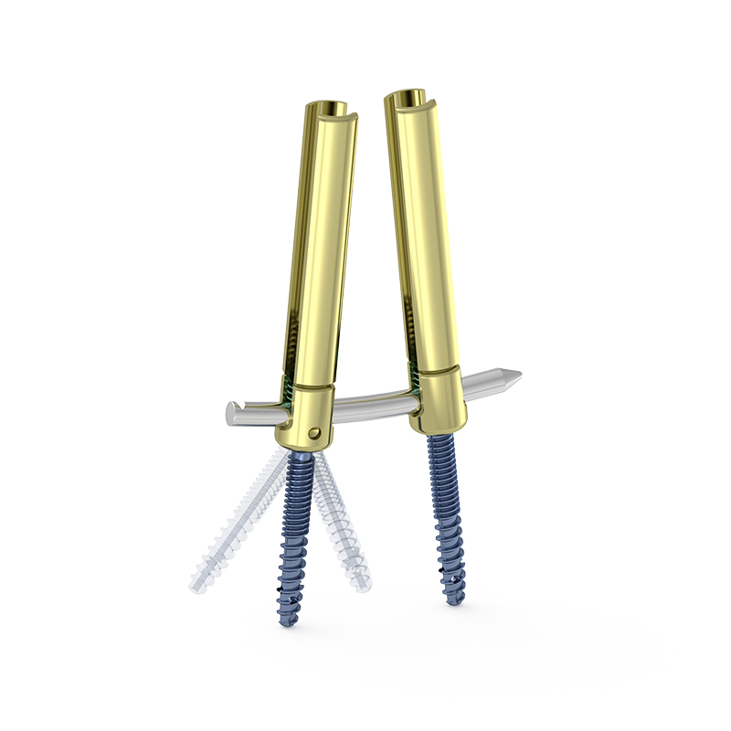
Pedicle screw placement is one of the most technically demanding steps in spinal surgery. When a surgeon positions each screw with precision, the entire construct gains stability, and the patient's recovery is protected from preventable complications. Even a small deviation in pedicle screw placement can have serious consequences, ranging from nerve damage to implant failure. Understanding why accurate pedicle screw placement is so critical helps surgeons, OR teams, and patients appreciate the high standards required in modern spine care.
The spine houses the spinal cord and a dense network of nerve roots, all of which lie in close proximity to the pedicle corridor used during pedicle screw placement. This anatomical reality makes accuracy non-negotiable. A well-executed pedicle screw placement secures vertebrae in proper alignment, supports fusion, and distributes mechanical load evenly across the construct. In contrast, a misplaced screw can compromise all of these goals simultaneously. This article explores the clinical, biomechanical, and patient-safety reasons why precise pedicle screw placement is essential in every spinal procedure.
Anatomy and the Risk Zones in Pedicle Screw Placement
The Narrow Corridor of the Pedicle
The pedicle is a small, cylindrical bridge of bone that connects the vertebral body to the posterior arch. Successful pedicle screw placement requires the screw to travel entirely within this corridor without breaching its walls. The medial wall of the pedicle is the most critical boundary, because a medial breach during pedicle screw placement places the screw in direct contact with the spinal cord or nerve roots. Even a breach of just two millimeters can produce neurological deficits that affect the patient permanently.
Lateral wall breaches during pedicle screw placement are also significant, as they can damage segmental vessels or leave the screw without adequate bone purchase. Inferior wall breaches may impinge on exiting nerve roots, causing radiculopathy. Each of these risks underscores why every step of pedicle screw placement must be planned and executed with anatomical respect and surgical precision.
Variation Across Spinal Levels
Pedicle screw placement becomes increasingly challenging in the thoracic spine, where pedicle diameters can be as narrow as four millimeters. At these levels, the margin for error in pedicle screw placement is almost zero. Surgeons must account for the natural angulation of the pedicle, the curvature of the spine, and any deformity present before committing to a trajectory. Advanced imaging and navigation technology have been developed specifically to improve the accuracy of pedicle screw placement at these difficult spinal levels.
Biomechanical Consequences of Inaccurate Pedicle Screw Placement
Construct Stability and Load Distribution
Every spinal fusion construct depends on pedicle screw placement that achieves solid cortical and cancellous bone purchase. When pedicle screw placement is accurate, the screw engages the dense bone of the pedicle and vertebral body, providing maximum pullout strength. If pedicle screw placement is off-axis or the screw exits the pedicle cortex, the fixation point becomes significantly weaker. This weakness can allow micromotion at the fusion site, which delays or prevents successful fusion and can lead to hardware loosening over time.
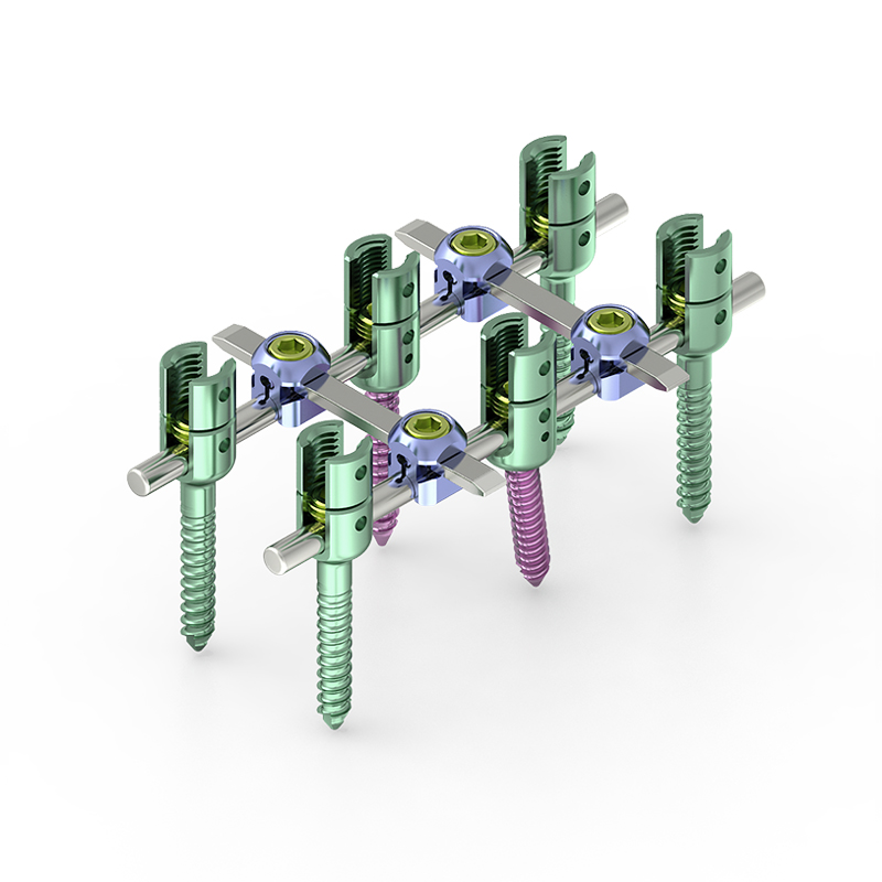
Inaccurate pedicle screw placement also disrupts the intended load-sharing mechanics of the spinal construct. When one screw is misplaced, the remaining screws must absorb disproportionate stress. This uneven load distribution accelerates fatigue in the hardware and increases the risk of rod fracture or screw breakage. Revision surgery for failed hardware is substantially more complex than the original procedure, making correct pedicle screw placement a strong factor in avoiding reoperation.
Alignment and Deformity Correction
In deformity correction cases, pedicle screw placement determines how effectively the surgeon can apply corrective forces to the spine. Reduction pedicle screws are specifically designed to facilitate this process by allowing the surgeon to reduce a displaced or angulated vertebra down to the rod. The effectiveness of reduction pedicle screw placement depends entirely on how accurately each screw has been inserted. If pedicle screw placement is inaccurate, the reduction maneuver may fail, leaving the deformity uncorrected or causing additional stress on surrounding structures.
Patient Safety Outcomes Linked to Pedicle Screw Placement Accuracy
Neurological Complications and Their Prevention
The most feared complication of inaccurate pedicle screw placement is neurological injury. Studies consistently show that the rate of symptomatic neurological complications is directly correlated with the accuracy of pedicle screw placement. Surgeons use multiple strategies to minimize this risk, including preoperative CT-based planning, intraoperative fluoroscopy, electrophysiological monitoring, and computer-assisted navigation. Each of these tools is designed to confirm that pedicle screw placement remains within the safe zone throughout the procedure.
Intraoperative neuromonitoring is particularly valuable during pedicle screw placement because it provides real-time feedback on neural function. If a signal change is detected during pedicle screw placement, the surgeon can immediately reassess the screw trajectory and correct it before permanent damage occurs. This level of vigilance reflects the critical importance that the surgical community places on accurate pedicle screw placement as a patient safety standard.
Long-Term Functional Recovery
Patients who undergo procedures with accurate pedicle screw placement typically experience better long-term functional outcomes. Solid fixation, achieved through precise pedicle screw placement, supports early mobilization and rehabilitation. Patients can begin physical therapy sooner, which reduces the risk of deconditioning, deep vein thrombosis, and prolonged hospital stays. Conversely, complications arising from inaccurate pedicle screw placement can delay rehabilitation for months and, in severe cases, result in permanent disability.
For patients undergoing spinal fusion with a pedicle screw placement system that includes reduction capability, the benefits extend to better coronal and sagittal alignment. Proper alignment achieved through accurate pedicle screw placement reduces adjacent segment stress and improves the durability of the surgical result over the patient's lifetime.
FAQ
What happens if pedicle screw placement is inaccurate?
Inaccurate pedicle screw placement can result in nerve injury, hardware failure, fusion nonunion, or the need for revision surgery. The severity depends on the direction and degree of the breach. Medial breaches are most dangerous because they risk direct contact with the spinal cord or nerve roots, while lateral or inferior breaches may cause vascular or radicular complications.
How do surgeons verify pedicle screw placement during surgery?
Surgeons verify pedicle screw placement using intraoperative fluoroscopy, cone-beam CT, or computer-assisted navigation systems. Electrophysiological monitoring with triggered electromyography is also used to detect any neural irritation caused by pedicle screw placement. These tools together provide a multi-layered confirmation that each screw is correctly positioned before the wound is closed.
What is a reduction pedicle screw and why does placement matter?
A reduction pedicle screw is a specialized implant designed with an extended tulip head that allows the surgeon to reduce a displaced vertebra onto the rod during the correction maneuver. The success of this technique depends entirely on accurate pedicle screw placement, because the reduction forces are transmitted through the screw-bone interface. If pedicle screw placement is imprecise, the reduction may fail or the screw may pull out under the corrective load.