Medical External Fixator System Solutions

All Categories

Get a Free Quote

Our representative will contact you soon.
Email
Name
Company Name
Message
0/1000

medical external fixator system

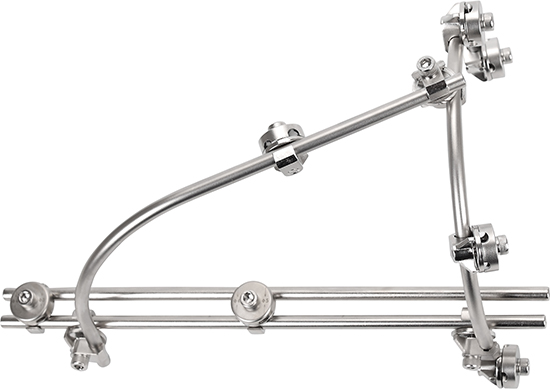
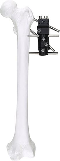
A medical external fixator system is an advanced orthopedic device designed to stabilize and support fractured or damaged bones from outside the body. This sophisticated system consists of pins, wires, or screws that are inserted through the skin into the bone, connected to an external frame made of carbon fiber, stainless steel, or aluminum alloy. The medical external fixator system plays a crucial role in modern trauma surgery and reconstructive orthopedics by providing reliable stabilization while allowing access to surrounding soft tissues for treatment. The primary functions include fracture stabilization, limb lengthening, deformity correction, and joint immobilization. Technologically, the medical external fixator system incorporates modular components that enable surgeons to customize configurations based on specific patient needs and injury patterns. Advanced designs feature multi-axial adjustment capabilities, radiolucent materials for clear radiographic imaging, and intuitive locking mechanisms for secure assembly. The system finds extensive applications in treating complex fractures, especially open fractures with severe soft tissue damage, pelvic ring disruptions, and cases requiring bone transport or gradual correction. The medical external fixator system is particularly valuable in emergency situations, polytrauma cases, and scenarios where internal fixation poses infection risks. Its versatility extends to pediatric orthopedics for growth-related corrections and in treating infected nonunions where hardware removal becomes necessary. With continuous technological refinements, the medical external fixator system remains an indispensable tool in contemporary orthopedic practice, offering surgeons flexibility and patients improved outcomes.

New Product Recommendations

The medical external fixator system delivers substantial practical benefits that directly impact patient recovery and surgical outcomes. One primary advantage is immediate fracture stabilization without requiring extensive surgical exposure, which significantly reduces operative time and minimizes tissue trauma. This approach proves especially beneficial when patients present with compromised soft tissue conditions or when swelling prevents immediate internal fixation. The system allows full access to wounds and surrounding tissues, enabling healthcare providers to perform regular dressing changes, monitor healing progress, and address soft tissue complications without removing stabilization hardware. From an operational perspective, the medical external fixator system offers remarkable adjustability, permitting post-operative modifications to alignment and compression without additional surgery. Surgeons can fine-tune bone positioning during the healing process, accommodating biological changes and optimizing fracture reduction. This adaptability translates to fewer revision procedures and improved final results. For facilities and practitioners, the system represents a cost-effective solution compared to multiple internal fixation attempts, particularly in contaminated or infected cases where implant retention becomes problematic. The medical external fixator system proves suitable across diverse applications, from temporary stabilization in damage control orthopedics to definitive treatment in complex reconstructions. Decision-makers appreciate its versatility in treating patients of all ages, from pediatric deformity corrections to geriatric fracture management. The system's modular design means institutions can maintain a comprehensive inventory that serves multiple clinical scenarios, maximizing resource utilization. Patients benefit from earlier mobilization potential, as the medical external fixator system often allows weight-bearing sooner than alternative treatments. The external positioning facilitates patient hygiene and comfort while reducing hospitalization duration, contributing to overall satisfaction and faster return to normal activities.

Tips And Tricks

How Do Pedicle Screw Systems Support Scoliosis Correction Surgery?

02

Jun

How Do Pedicle Screw Systems Support Scoliosis Correction Surgery?

Scoliosis correction surgery is one of the most technically demanding procedures in modern spinal orthopedics. The ability to realign a severely curved spine, restore sagittal balance, and maintain long-term structural integrity depends heavily on th...
View More
What Are the Indications for Using Pedicle Screw in Degenerative Spine Disease?

02

Jun

What Are the Indications for Using Pedicle Screw in Degenerative Spine Disease?

Degenerative spine disease encompasses a broad spectrum of conditions that progressively compromise spinal stability, alignment, and neurological function. As these conditions advance, conservative management often fails to provide adequate relief, a...
View More
How Does Pedicle Screw Design Enhance Pullout Strength and Stability?

02

Jun

How Does Pedicle Screw Design Enhance Pullout Strength and Stability?

In modern spinal surgery, fixation reliability is not a secondary consideration — it is the foundation upon which patient outcomes are built. The pedicle screw has become the cornerstone of posterior spinal instrumentation, offering surgeons a ...
View More
Why Is Pedicle Screw Accuracy Critical in Minimally Invasive Spine Surgery?

02

Jun

Why Is Pedicle Screw Accuracy Critical in Minimally Invasive Spine Surgery?

In the evolving landscape of spinal surgery, precision is not merely a technical preference — it is a clinical imperative. The pedicle screw has become the cornerstone of modern spinal fixation systems, offering surgeons a reliable anchor point...
View More

Get a Free Quote

Our representative will contact you soon.
Email
Name
Company Name
Message
0/1000

medical external fixator system

Advanced Modular Configuration for Customized Treatment

Advanced Modular Configuration for Customized Treatment

The medical external fixator system features an innovative modular design that empowers surgeons to create patient-specific configurations tailored to unique anatomical requirements and injury patterns. This modularity encompasses interchangeable rings, bars, connectors, and fixation elements that can be assembled in virtually unlimited combinations. Surgeons can select from various frame geometries including circular, hybrid, and unilateral constructs depending on fracture location, bone quality, and treatment objectives. The system accommodates both Ilizarov-type circular frames for comprehensive three-dimensional correction and simpler rail-based designs for straightforward stabilization needs. Each component integrates seamlessly with others through standardized connection interfaces, ensuring mechanical stability while maintaining adjustment flexibility. This adaptability proves invaluable when addressing complex deformities requiring gradual correction over extended periods, as the medical external fixator system can be modified incrementally without complete reconstruction. The modular approach also benefits healthcare facilities by reducing inventory complexity, as a core set of universal components serves multiple clinical applications rather than requiring procedure-specific kits. This design philosophy translates to more efficient surgical planning, reduced setup time in operating rooms, and enhanced ability to respond to intraoperative findings that may necessitate configuration changes.
Radiolucent Materials for Superior Imaging Clarity

Radiolucent Materials for Superior Imaging Clarity

Modern iterations of the medical external fixator system incorporate advanced radiolucent materials, particularly carbon fiber composites, that dramatically improve radiographic visualization during and after treatment. Traditional metal frames often create significant imaging artifacts that obscure fracture lines, callus formation, and alignment assessment, complicating clinical decision-making. The integration of radiolucent components eliminates these obstacles, providing surgeons with unobstructed views of the treatment area during fluoroscopic guidance and follow-up radiographs. This technological advancement enables more accurate initial fracture reduction, as surgeons can verify positioning without frame interference during surgical application. Throughout the healing process, healthcare providers can monitor bone consolidation, detect complications early, and determine optimal timing for frame removal based on clear imaging evidence. The medical external fixator system utilizing radiolucent materials also facilitates CT and MRI compatibility in cases requiring advanced imaging, expanding diagnostic capabilities without hardware removal. Beyond imaging benefits, carbon fiber components offer excellent strength-to-weight ratios, reducing the overall system mass that patients must bear while maintaining structural integrity comparable to steel alternatives. This weight reduction contributes to improved patient comfort, easier ambulation, and decreased muscle fatigue during extended treatment periods. The combination of imaging transparency and mechanical performance positions radiolucent medical external fixator system technology as a significant advancement over conventional metallic designs.
Multi-Axial Adjustment Capability for Precise Correction

Multi-Axial Adjustment Capability for Precise Correction

The medical external fixator system distinguishes itself through sophisticated multi-axial adjustment mechanisms that enable precise three-dimensional corrections throughout treatment duration. Unlike static fixation devices, this system incorporates specialized hinges, translation units, and telescoping struts that permit controlled movements in multiple planes simultaneously. Surgeons can program gradual corrections for angulation, rotation, translation, and length discrepancies by making systematic adjustments according to prescribed protocols. This capability proves essential in treating complex deformities where correction must occur progressively to allow soft tissue adaptation and avoid neurovascular complications from abrupt realignment. The medical external fixator system typically features calibrated adjustment mechanisms with clear measurement scales, ensuring reproducible and accurate corrections at each adjustment interval. Patients or caregivers can often perform prescribed adjustments at home following proper training, reducing clinic visits while maintaining treatment progression. This home-based adjustment capability increases treatment accessibility for patients in remote locations and reduces healthcare system burden. The multi-axial functionality also addresses unexpected complications, as surgeons can modify treatment plans dynamically if initial correction strategies prove insufficient or if patients develop secondary issues. Digital planning software now integrates with many medical external fixator system platforms, allowing preoperative virtual simulations that guide frame application and adjustment schedules, further enhancing precision and predictability in complex reconstructive cases.

Get a Free Quote

Our representative will contact you soon.
Email
Name
Company Name
Message
0/1000
Newsletter
Please Leave A Message With Us